Fraud Blocker Skip to main content

Anatomy Collection (page 18)

"Unveiling the Intricacies of Anatomy: From Sensory Homunculus to Motor Homunculus" Step into the fascinating world of anatomy, where every detail tells a story

Background imageAnatomy Collection: A woodcut engraving depicting a blood-letting man, showing points of blood-letting connected with

A woodcut engraving depicting a blood-letting man, showing points of blood-letting connected with signs of the Zodiac
5311213 A woodcut engraving depicting a blood-letting man, showing points of blood-letting connected with signs of the Zodiac, 16th century; Universal History Archive/UIG

Background imageAnatomy Collection: Diagram of the circulation of blood as understood after William Harvey's work

Diagram of the circulation of blood as understood after William Harvey's work
5309470 Diagram of the circulation of blood as understood after William Harvey's work, showing the Lesser Circulation (Pulmonary Circulation)

Background imageAnatomy Collection: Direct sphygmograph, 1882 (engraving)

Direct sphygmograph, 1882 (engraving)
8766934 Direct sphygmograph, 1882 (engraving); Private Collection; (add.info.: Direct sphygmograph. Dr. Jaccoud-V 33 1882); Photo ©Jaime Abecasis

Background imageAnatomy Collection: Crossing section of fibro-nucleated tumors, 1867 (engraving)

Crossing section of fibro-nucleated tumors, 1867 (engraving)
8766909 Crossing section of fibro-nucleated tumors, 1867 (engraving); Private Collection; (add.info.: Crossing section of fibro-nucleated tumors. ( Dr. Jaccoud dictionary.Vol)

Background imageAnatomy Collection: Dog Skulls and Skeleton Set - Dogs skulls and skeleton, Museum d'histoire naturelle de Marseille

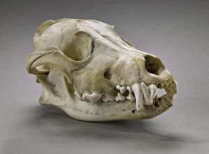
Dog Skulls and Skeleton Set - Dogs skulls and skeleton, Museum d'histoire naturelle de Marseille
CSO4987875 Dog Skulls and Skeleton Set - Dogs skulls and skeleton, Museum d'histoire naturelle de Marseille; (add.info.: Dog Skulls and Skeleton Set - Dogs skulls and skeleton)

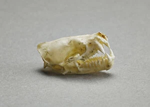
Background imageAnatomy Collection: Talpa Europea (Skeleton of an European mole), Museum d'histoire naturelle de Marseille

Talpa Europea (Skeleton of an European mole), Museum d'histoire naturelle de Marseille
CSO4987854 Talpa Europea (Skeleton of an European mole), Museum d'histoire naturelle de Marseille; (add.info.: Talpa Europea (Skeleton of an European mole)

Background imageAnatomy Collection: European or noble crayfish or crawfish (ecrevisse a pate rouge), Astacus astacus. Vulnerable

European or noble crayfish or crawfish (ecrevisse a pate rouge), Astacus astacus. Vulnerable
FLO4575853 European or noble crayfish or crawfish (ecrevisse a pate rouge), Astacus astacus. Vulnerable. Crayfish 1, claws 2, 3, eye 4, internal organs 5, and jaw muscle 6

Background imageAnatomy Collection: Anatomy of a tree: cross section of pine branch 1b, wood chip 1a, the same magnified 2

Anatomy of a tree: cross section of pine branch 1b, wood chip 1a, the same magnified 2, magnified further 3
FLO4574347 Anatomy of a tree: cross section of pine branch 1b, wood chip 1a, the same magnified 2, magnified further 3. Handcoloured copperplate engraving from Bertuch's " Bilderbuch fur

Background imageAnatomy Collection: The entrails in the human body: lungs, liver, diaphragm, stomach, duodenum, colon, etc

The entrails in the human body: lungs, liver, diaphragm, stomach, duodenum, colon, etc
FLO4574696 The entrails in the human body: lungs, liver, diaphragm, stomach, duodenum, colon, etc. Handcoloured copperplate engraving from Bertuch's " Bilderbuch fur Kinder"

Background imageAnatomy Collection: Human airways 1866 illustration of the voice box (larynx, top), trachea (windpipe, vertical)

Human airways 1866 illustration of the voice box (larynx, top), trachea (windpipe, vertical)
JAB4119233 Human airways 1866 illustration of the voice box (larynx, top), trachea (windpipe, vertical, centre) and bronchi (lower left and right)

Background imageAnatomy Collection: Human skull 1866 illustration showing the anatomy of the front (Fig 1) and rear (Fig 2)

Human skull 1866 illustration showing the anatomy of the front (Fig 1) and rear (Fig 2)
JAB4119205 Human skull 1866 illustration showing the anatomy of the front (Fig 1) and rear (Fig 2) of a human skull Le crane - Fig 1: Facial region: frontal bone, frontal bump, eyebrow arcade

Background imageAnatomy Collection: Circulatory system. (La vie normale et la vie normale de J.Rengade - lymph nodes du cou et la)

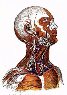
Circulatory system. (La vie normale et la vie normale de J.Rengade - lymph nodes du cou et la aisselle.Drawing A)
JAB4120095 Circulatory system. (La vie normale et la vie normale de J.Rengade - lymph nodes du cou et la aisselle.Drawing A. Demarle 1881; (add.info.: Circulatory system)

Background imageAnatomy Collection: Page of a manuscript by Pedanius Dioscorides (Dioscorides) Anazarebus, Greek physician. 13th century

Page of a manuscript by Pedanius Dioscorides (Dioscorides) Anazarebus, Greek physician. 13th century
GIA5036777 Page of a manuscript by Pedanius Dioscorides (Dioscorides) Anazarebus, Greek physician. 13th century.; © Giancarlo Costa

Background imageAnatomy Collection: Portrait of Wilhelm Roux (1850-1924) German anatomist

Portrait of Wilhelm Roux (1850-1924) German anatomist
XEE5031769 Portrait of Wilhelm Roux (1850-1924) German anatomist; (add.info.: Portrait of Wilhelm Roux (1850-1924) German anatomist); Stefano Bianchetti

Background imageAnatomy Collection: Le Coeur in ' Treats on the structure of the heart, its action, and its diseases'

Le Coeur in " Treats on the structure of the heart, its action, and its diseases"
XEE5031961 Le Coeur in " Treats on the structure of the heart, its action, and its diseases", 1749 by Jean Baptiste SENAC (1693-1770); (add.info)

Background imageAnatomy Collection: Portrait of Nikolai Ivanovich Pirogov (1810-1881) Russian anatomist and surgeon

Portrait of Nikolai Ivanovich Pirogov (1810-1881) Russian anatomist and surgeon
XEE5031004 Portrait of Nikolai Ivanovich Pirogov (1810-1881) Russian anatomist and surgeon; (add.info.: Portrait of Nikolai Ivanovich Pirogov (1810-1881) Russian anatomist and surgeon

Background imageAnatomy Collection: Portrait of Francis Glisson (1597 1677) British physician, anatomist, physiologist, and pathologist

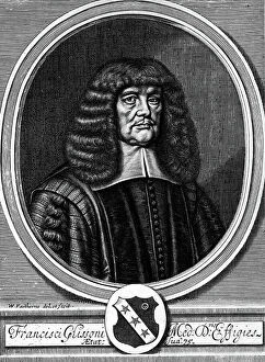
Portrait of Francis Glisson (1597 1677) British physician, anatomist, physiologist, and pathologist
XEE5029034 Portrait of Francis Glisson (1597 1677) British physician, anatomist, physiologist, and pathologist, engraving 18th century; (add.info)

Background imageAnatomy Collection: Portrait of JEAN CRUVEILHIER (1791-1874), French anatomist

Portrait of JEAN CRUVEILHIER (1791-1874), French anatomist
XEE5028654 Portrait of JEAN CRUVEILHIER (1791-1874), French anatomist; (add.info.: Portrait of JEAN CRUVEILHIER (1791-1874), French anatomist); Stefano Bianchetti

Background imageAnatomy Collection: Canis lupus (Wolf gris - gray (grey) woolf or timber woolf), Museum d'histoire naturelle de

Canis lupus (Wolf gris - gray (grey) woolf or timber woolf), Museum d'histoire naturelle de Marseille
CSO5006426 Canis lupus (Wolf gris - gray (grey) woolf or timber woolf), Museum d'histoire naturelle de Marseille; (add.info.: Canis lupus (Wolf gris - gray (grey) woolf or timber woolf)

Background imageAnatomy Collection: Frontispice of ' Exercisitationes anatomicae duae de structura et sus renum' of 1725 by Lorenzo

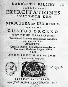
Frontispice of " Exercisitationes anatomicae duae de structura et sus renum" of 1725 by Lorenzo (Laurentio)
XEE5028108 Frontispice of " Exercisitationes anatomicae duae de structura et sus renum" of 1725 by Lorenzo (Laurentio) Bellini (Firenze 1643 - Pisa 1704) - Biblioteca nazionale braidense

Background imageAnatomy Collection: Portrait of Bernard Siegfried Albinus (1697-1770) German anatomist

Portrait of Bernard Siegfried Albinus (1697-1770) German anatomist
XEE5027659 Portrait of Bernard Siegfried Albinus (1697-1770) German anatomist; (add.info.: Portrait of Bernard Siegfried Albinus (1697-1770) German anatomist); Stefano Bianchetti

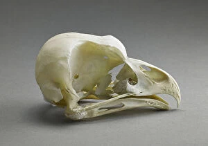
Background imageAnatomy Collection: Bubo bubo (Skull of the Eurasian eagle-owl), Museum of Natural History of Marseille

Bubo bubo (Skull of the Eurasian eagle-owl), Museum of Natural History of Marseille
CSO5006276 Bubo bubo (Skull of the Eurasian eagle-owl), Museum of Natural History of Marseille; (add.info.: Bubo bubo (Skull of the Eurasian eagle-owl)

Background imageAnatomy Collection: Vesperugo serotinus (Chiroptere) (Crane de la grande serotine ou serotine commune - skull of an)

Vesperugo serotinus (Chiroptere) (Crane de la grande serotine ou serotine commune - skull of an european long-eared bat)
CSO5006273 Vesperugo serotinus (Chiroptere) (Crane de la grande serotine ou serotine commune - skull of an european long-eared bat or European brown bat)

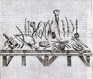
Background imageAnatomy Collection: Instrument surgical csurgical instruments) - in ' De anatomome corporis humani' by Guido Guidi

Instrument surgical csurgical instruments) - in " De anatomome corporis humani" by Guido Guidi (Vidus Vidius)
XEE5029681 Instrument surgical csurgical instruments) - in " De anatomome corporis humani" by Guido Guidi (Vidus Vidius) (1509-1569) Italian surgeon and anatomist

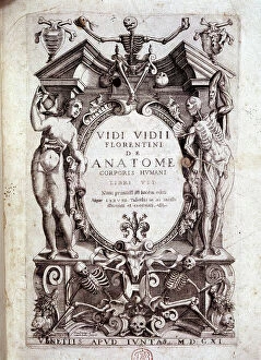
Background imageAnatomy Collection: Frontispice of ' De anatomome corporis humani' by Guido Guidi (Vidus Vidius) (1509-1569)

Frontispice of " De anatomome corporis humani" by Guido Guidi (Vidus Vidius) (1509-1569)
XEE5029679 Frontispice of " De anatomome corporis humani" by Guido Guidi (Vidus Vidius) (1509-1569) Italian surgeon and anatomist

Background imageAnatomy Collection: Anatomy: representation of the uterus by Garioponto (970 - 1050) Italian doctor of the Middle Ages

Anatomy: representation of the uterus by Garioponto (970 - 1050) Italian doctor of the Middle Ages
XEE5029571 Anatomy: representation of the uterus by Garioponto (970 - 1050) Italian doctor of the Middle Ages, Biblioteca Civica Bertoliana

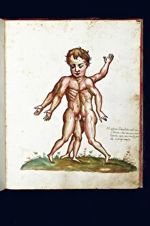
Background imageAnatomy Collection: Monster creature. Illustrated chart of a manuscript of Natural History by Ulisse Aldrovandi

Monster creature. Illustrated chart of a manuscript of Natural History by Ulisse Aldrovandi (1522-1605)
VEN5029506 Monster creature. Illustrated chart of a manuscript of Natural History by Ulisse Aldrovandi (1522-1605), Ulisse Aldrovandi fonds, University Library of Bologna, ed

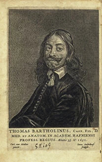
Background imageAnatomy Collection: BARTHOLIN, Thomas (1616-1680). Danish physician, mathematician, and theologian

BARTHOLIN, Thomas (1616-1680). Danish physician, mathematician, and theologian
CUL4847818 BARTHOLIN, Thomas (1616-1680). Danish physician, mathematician, and theologian. He is best known for his work in the discovery of the lymphatic system in humans

Background imageAnatomy Collection: Front view of a healthy female pelvis. Copperplate engraving by Andrew Bell after an illustration

Front view of a healthy female pelvis. Copperplate engraving by Andrew Bell after an illustration by Jan van Rymsdyk
FLO4695855 Front view of a healthy female pelvis. Copperplate engraving by Andrew Bell after an illustration by Jan van Rymsdyk from William Smellie's A Set of Anatomical Tables, Charles Elliot

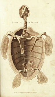
Background imageAnatomy Collection: Skeleton of a turtle. Handcoloured copperplate engraving by Heath after an illustration by George

Skeleton of a turtle. Handcoloured copperplate engraving by Heath after an illustration by George Shaw from his General
FLO4694475 Skeleton of a turtle. Handcoloured copperplate engraving by Heath after an illustration by George Shaw from his General Zoology, Amphibia, London, 1801.; (add.info.: Skeleton of a turtle)

Background imageAnatomy Collection: La scarlatine ou feevre ecarlate - Lithograph by John Pass, published in 'Encyclopedia

La scarlatine ou feevre ecarlate - Lithograph by John Pass, published in "Encyclopedia Londinensis" by John
FLO4618755 La scarlatine ou feevre ecarlate - Lithograph by John Pass, published in "Encyclopedia Londinensis" by John Wilkes, edition J.Adlard, London

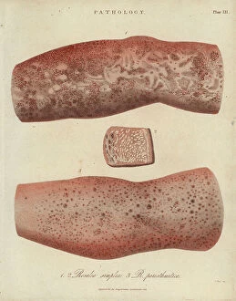
Background imageAnatomy Collection: Skin Diseases: Nodular or Hyde Prurigo and Varietes of Psoriasis - Lithography by John Pass

Skin Diseases: Nodular or Hyde Prurigo and Varietes of Psoriasis - Lithography by John Pass
FLO4618824 Skin Diseases: Nodular or Hyde Prurigo and Varietes of Psoriasis - Lithography by John Pass, published in "Encyclopedia Londinensis" by John Wilkes, J.Adlard Edition, London

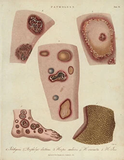
Background imageAnatomy Collection: Ichthyosis (peeling), dyshidrosis (bullous eczema) and herpetic skin diseases -Lithography by John

Ichthyosis (peeling), dyshidrosis (bullous eczema) and herpetic skin diseases -Lithography by John Pass
FLO4618812 Ichthyosis (peeling), dyshidrosis (bullous eczema) and herpetic skin diseases -Lithography by John Pass, published in "Encyclopedia Londinensis" by John Wilkes, J.Adlard edition

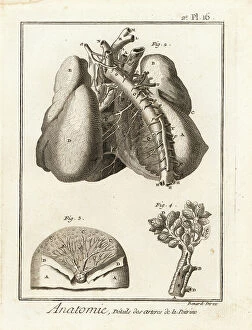
Background imageAnatomy Collection: Arteries in the chest cavity to the heart and lungs. Copperplate engraving by Robert Benard after

Arteries in the chest cavity to the heart and lungs. Copperplate engraving by Robert Benard after an illustration by
FLO4611670 Arteries in the chest cavity to the heart and lungs. Copperplate engraving by Robert Benard after an illustration by Albrecht von Haller from Denis Diderot's Encyclopedia, Pellet

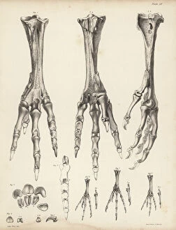
Background imageAnatomy Collection: Metatarsus and toes of the dodo, Raphus cucullatus 1-5, foot of the yellow-footed green pigeon

Metatarsus and toes of the dodo, Raphus cucullatus 1-5, foot of the yellow-footed green pigeon, Treron chlorigaster 6
FLO4612178 Metatarsus and toes of the dodo, Raphus cucullatus 1-5, foot of the yellow-footed green pigeon, Treron chlorigaster 6, stock dove, Columba oenas 7 and partridge pigeon

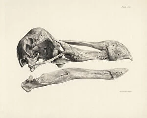
Background imageAnatomy Collection: Side view of the skull of a dodo. Lithograph from Hugh Edwin Strickland

Side view of the skull of a dodo. Lithograph from Hugh Edwin Strickland and Alexander Gordon Melville's The Dodo
FLO4612152 Side view of the skull of a dodo. Lithograph from Hugh Edwin Strickland and Alexander Gordon Melville's The Dodo and its Kindred, London, Reeve, Benham and Reeve

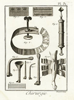
Background imageAnatomy Collection: Sutures, withers, fine-tip syringe by Dominique Anel, siphon, injection tubes

Sutures, withers, fine-tip syringe by Dominique Anel, siphon, injection tubes
FLO4611953 Sutures, withers, fine-tip syringe by Dominique Anel, siphon, injection tubes, 18-sheet bandage for complex fractures

Background imageAnatomy Collection: France: Medical photograph of a spinal malformation, 1890

France: Medical photograph of a spinal malformation, 1890
LUX4755402 France: Medical photograph of a spinal malformation, 1890; (add.info.: France: Medical photograph of a spinal malformation, 1890); Photo © Lux-in-Fine

Background imageAnatomy Collection: Bertillonage (Bertillon system or forensic anthropometry) a forensic technique developed by

Bertillonage (Bertillon system or forensic anthropometry) a forensic technique developed by the Frenchman Alphonse
ELD4960696 Bertillonage (Bertillon system or forensic anthropometry) a forensic technique developed by the Frenchman Alphonse Bertillon (1853-1914) in 1879

Background imageAnatomy Collection: Anatomy of the human skeleton, 1820 (copperplate engraving)

Anatomy of the human skeleton, 1820 (copperplate engraving)
FLO4591008 Anatomy of the human skeleton, 1820 (copperplate engraving); (add.info.: Anatomy of the human skeleton. Copperplate engraving by Milton from Abraham Rees Cyclopedia or Universal Dictionary)

Background imageAnatomy Collection: Inca Crane proving the practice of surgical operation on the human brain during prehistory in Peru

Inca Crane proving the practice of surgical operation on the human brain during prehistory in Peru Colour engraving of
NWI4900256 Inca Crane proving the practice of surgical operation on the human brain during prehistory in Peru Colour engraving of the 19th century; (add.info)

Background imageAnatomy Collection: Sperm whales in ' Painting Encyclopedique et Methodique des trois Regnes de la Nature' by Pierre

Sperm whales in " Painting Encyclopedique et Methodique des trois Regnes de la Nature" by Pierre Joseph
FAT4299955 Sperm whales in " Painting Encyclopedique et Methodique des trois Regnes de la Nature" by Pierre Joseph Bonnaterre (1762-1804)

Background imageAnatomy Collection: Abdominal muscles - 1866 illustration showing the structure of the muscles (brown)

Abdominal muscles - 1866 illustration showing the structure of the muscles (brown) and tendons (white)
JAB4119475 Abdominal muscles - 1866 illustration showing the structure of the muscles (brown) and tendons (white) found in the front of the human abdomen - Region abdominale anterieure - Muscle

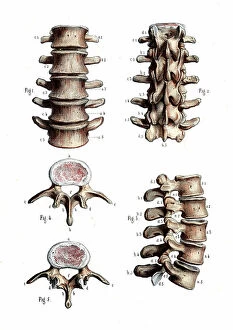
Background imageAnatomy Collection: Lumbar spine anatomy - 1866 anatomical illustration of the lumbar spine (lower back)

Lumbar spine anatomy - 1866 anatomical illustration of the lumbar spine (lower back) - Lumbar vertebrae
JAB4119471 Lumbar spine anatomy - 1866 anatomical illustration of the lumbar spine (lower back) - Lumbar vertebrae, vertebral column - Osteology, locomotion

Background imageAnatomy Collection: Chest and shoulder muscles - 1866 anatomical illustration showing the structure of the muscles

Chest and shoulder muscles - 1866 anatomical illustration showing the structure of the muscles (brown)
JAB4119460 Chest and shoulder muscles - 1866 anatomical illustration showing the structure of the muscles (brown) and tendons (white) of the rear of the upper back

Background imageAnatomy Collection: Foot anatomy - 1866 anatomical illustration showing the muscles (brown)

Foot anatomy - 1866 anatomical illustration showing the muscles (brown), tendons (white) and bones (beige)
JAB4119455 Foot anatomy - 1866 anatomical illustration showing the muscles (brown), tendons (white) and bones (beige) of the human foot - Plantaire region- Anatomy - Locomotion apparatus, myology

Background imageAnatomy Collection: Human hand - 1866 anatomical illustration showing the muscles (brown) and tendons (white)

Human hand - 1866 anatomical illustration showing the muscles (brown) and tendons (white)
JAB4119450 Human hand - 1866 anatomical illustration showing the muscles (brown) and tendons (white) of the human hand - Region palmar- Anatomy - Fig 1 Cubital anterieur Fig 2 cubital anterieur et

Background imageAnatomy Collection: Leg joints - 1866 anatomical illustration of the muscles (brown), tendons (white) and bones (beige)

Leg joints - 1866 anatomical illustration of the muscles (brown), tendons (white) and bones (beige)
JAB4119440 Leg joints - 1866 anatomical illustration of the muscles (brown), tendons (white) and bones (beige) of the human hip (top) and knee (bottom) joints


All Professionally Made to Order for Quick Shipping

"Unveiling the Intricacies of Anatomy: From Sensory Homunculus to Motor Homunculus" Step into the fascinating world of anatomy, where every detail tells a story. Explore the depths of human and animal structures as we delve into this captivating subject. Let's begin with Leonardo da Vinci's masterpiece, "Head of a Bear. " Created in 1480 but discovered in 1945, this artwork showcases da Vinci's unparalleled ability to capture anatomical accuracy. The intricate details reveal his keen observation skills and deep understanding of form. Moving on to our furry friends, the greyhound takes center stage. Discovering the anatomy of these majestic creatures unveils their remarkable speed and agility. From their sleek skeleton to their muscular build, it is evident why they are renowned for their racing abilities. But what about us humans? Enter the sensory homunculus – a visual representation that depicts how our brain perceives different body parts based on sensitivity levels. This map reveals intriguing insights into how our senses are distributed throughout our bodies. X-ray images provide another dimension to understanding anatomy. Take a glimpse at normal knees through an X-ray lens – marvel at the complexity hidden beneath our skin while appreciating its delicate balance between strength and flexibility. The backbone is often considered one of nature's most ingenious designs. Our human backbone includes ribs and pelvis, providing stability while allowing movement – truly an architectural marvel worth exploring further. Venturing deeper within ourselves, let us explore the intricacies of the human brain from an inferior view. Witness its complex network of connections responsible for controlling various bodily functions - truly awe-inspiring. As we journey back in time, we encounter hominid crania – remnants that shed light on our evolutionary history. These ancient skulls offer glimpses into early forms such as Australopithecus afarensis (AL 288-1), famously known as Lucy - bridging gaps between past and present.