Fraud Blocker Skip to main content

Anatomy Collection (page 6)

"Unveiling the Intricacies of Anatomy: From Sensory Homunculus to Motor Homunculus" Step into the fascinating world of anatomy, where every detail tells a story

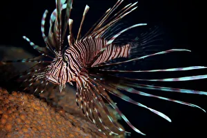
Background imageAnatomy Collection: Red Lionfish flares its deadly spines

Red Lionfish flares its deadly spines as a warning to the photographer not to get any closer, Bonaire, Caribbean Netherlands

Background imageAnatomy Collection: The eye according to Hunain ibn Ishaq also called Johannitius (809-873) Baghdad physican

The eye according to Hunain ibn Ishaq also called Johannitius (809-873) Baghdad physican and philosopher. Manuscript c1200

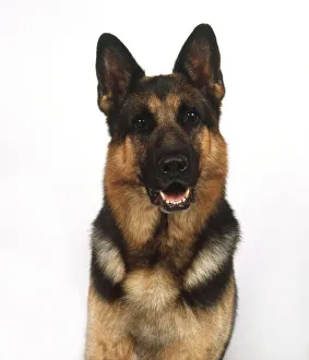
Background imageAnatomy Collection: Head and shoulders of a German Shepherd (Canis familiaris), front view

Head and shoulders of a German Shepherd (Canis familiaris), front view

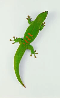
Background imageAnatomy Collection: Madagascan Giant Day Gecko (Gekkonidae), bright green with red markings, view from above

Madagascan Giant Day Gecko (Gekkonidae), bright green with red markings, view from above

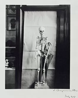
Background imageAnatomy Collection: Skeleton of Negrillo or pigmy

Skeleton of Negrillo or pigmy
A skeleton of a female pygmy from the Akka Tribe, Monnattu, central Africa. Specimen presented by Dr Emin Pasha, 1887. Photograph by J Benjamin Stone in 1907

Background imageAnatomy Collection: Tyrannosaurus rex skeleton

Tyrannosaurus rex skeleton
A skeleton of the carnivorous dinosaur, Tyrannosaurus rex at the Natural History Museum. The Tyrannosaurus rex, which grew up to around 12 metres long lived 67 to 65 million years ago during

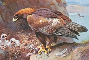
Background imageAnatomy Collection: Aquila chrysaetus, golden eagle

Aquila chrysaetus, golden eagle
Plate 78 from Archibald Thorburns second edtition of British Birds, Vol. 2 (1925)

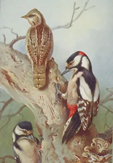
Background imageAnatomy Collection: Jynx torquilla, wryneck, Dendrocopos major, great spotted wo

Jynx torquilla, wryneck, Dendrocopos major, great spotted wo
Plate 60 from Archibald Thorburns second edtition of British Birds, Vol. 2 (1925)

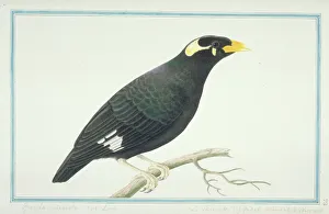
Background imageAnatomy Collection: Gracula religiosa indica, lesser hill myna

Gracula religiosa indica, lesser hill myna
Plate 73, painting by Pieter Cornelius de Bevere, from the Loten Collection of coloured drawings of Birds, Mammals, Insects & Plants, (1754-57)

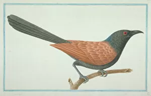
Background imageAnatomy Collection: Centropus sinensis, greater coucal

Centropus sinensis, greater coucal
Plate 27, painting by Pieter Cornelius de Bevere, from the Loten Collection of coloured drawings of Birds, Mammals, Insects & Plants, (1754-57)

Background imageAnatomy Collection: Gavia stellata, red-throated diver

Gavia stellata, red-throated diver
Plate 45 from John Goulds The Birds of Great Britain, Vol. 5 (1873). Hand coloured lithograph

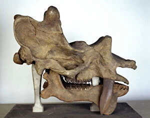
Background imageAnatomy Collection: Uintatherium skull

Uintatherium skull
Skull measures 740 mm left to right. Uintatherium, a horned ungulate from the mid Eocene of western U.S.A, stood about 1.6m at the shoulder
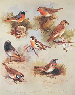
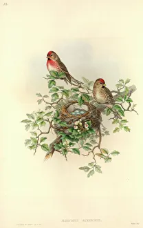
Background imageAnatomy Collection: Carduelis flammea cabaret, lesser redpoll

Carduelis flammea cabaret, lesser redpoll
Plate 52 from John Goulds The Birds of Great Britain, Vol. 3 (1873). Hand coloured lithograph
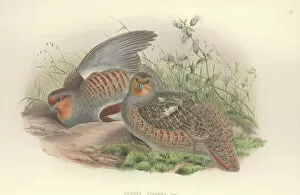
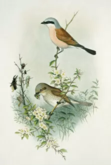
Background imageAnatomy Collection: Lanius collurio, red-backed shrike

Lanius collurio, red-backed shrike
Plate 15 from John Goulds The Birds of Great Britain, Vol. 2 (1873). Hand coloured lithograph

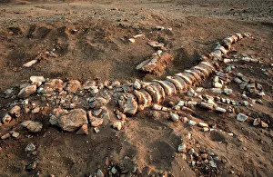
Background imageAnatomy Collection: Dinosaur tail bones

Dinosaur tail bones
From a Palaeontology field trip in Niger, West Africa

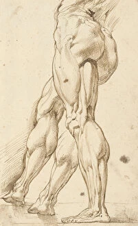
Background imageAnatomy Collection: An ecorche study of the legs of a male nude, with a subsidiary study of the right leg

An ecorche study of the legs of a male nude, with a subsidiary study of the right leg
3588687 An ecorchestudy of the legs of a male nude, with a subsidiary study of the right leg (pen & brown ink on paper) by Rubens

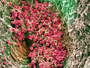
Background imageAnatomy Collection: Anatomy, Biology, Blood Vessel, Cell, Color Image, Fibrin, Healthcare And Medicine

Anatomy, Biology, Blood Vessel, Cell, Color Image, Fibrin, Healthcare And Medicine
Anatomy, Biology, Blood Vessel, Cell, Color Image, Fibrin, Healthcare, Science Photo Library, 85758208

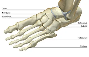
Background imageAnatomy Collection: above view, anatomy, bone, bone structure, bone structure of the foot, bones, bones of the foot

above view, anatomy, bone, bone structure, bone structure of the foot, bones, bones of the foot
above view, anatomy, bone, bone structure, bone structure of the foo, Medical Image Collection, 87396631

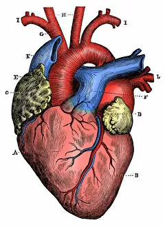
Background imageAnatomy Collection: Antique medical scientific illustration high-resolution: heart

Antique medical scientific illustration high-resolution: heart

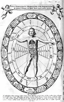
Background imageAnatomy Collection: Influence of the Universe, the Macrocosm, on Man, the Microcosm, 1678

Influence of the Universe, the Macrocosm, on Man, the Microcosm, 1678. From Mundus Subterraneus by Athanasius Kircher. (Amsterdam, 1678)

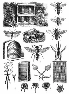
Background imageAnatomy Collection: Beekeeping

Beekeeping (or apiculture, from Latin: apis bee ) is the maintenance of honey bee colonies, commonly in hives

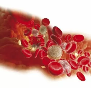
Background imageAnatomy Collection: Diagram showing bloodstream inside a vein, red, white blood cells, platelet

Diagram showing bloodstream inside a vein, red, white blood cells, platelet

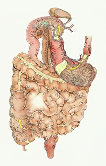
Background imageAnatomy Collection: Human digestive system

Human digestive system

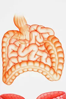
Background imageAnatomy Collection: Illustration of human digestive system, including stomach, small intestine and large intestine

Illustration of human digestive system, including stomach, small intestine and large intestine

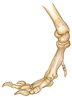
Background imageAnatomy Collection: Illustration of bones of Tyrannosaurus foot showing long toes, small first toe, or dew claw

Illustration of bones of Tyrannosaurus foot showing long toes, small first toe, or dew claw, and ankle joint

Background imageAnatomy Collection: Digital cross section illustration of taste buds and filiform papilae on tongue

Digital cross section illustration of taste buds and filiform papilae on tongue

Background imageAnatomy Collection: Cross section biomedical illustration of iliac crest site on pelvis

Cross section biomedical illustration of iliac crest site on pelvis

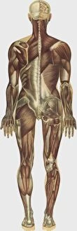
Background imageAnatomy Collection: The human body with superimposed colored plates by Julien Bougle

The human body with superimposed colored plates by Julien Bougle
The human body with superimposed colored plates, by Julien Bougle, circa 1899

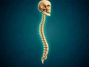
Background imageAnatomy Collection: Conceptual image of human skull and spinal cord

Conceptual image of human skull and spinal cord

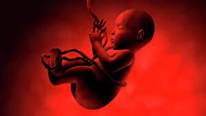
Background imageAnatomy Collection: Human fetus inside amniotic sac

Human fetus inside amniotic sac

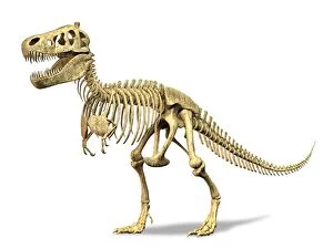
Background imageAnatomy Collection: 3D rendering of a Tyrannosaurus Rex dinosaur skeleton

3D rendering of a Tyrannosaurus Rex dinosaur skeleton. T-Rex was one of the largest carnivorous dinosaurs of the Cretaceous period

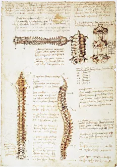
Background imageAnatomy Collection: Pen and ink studies of the human spinal column by Leonardo da Vinci, c1510-11

Pen and ink studies of the human spinal column by Leonardo da Vinci, c1510-11
LEONARDO: ANATOMY, c1510. Pen and ink studies of the human spinal column by Leonardo da Vinci, c1510-11

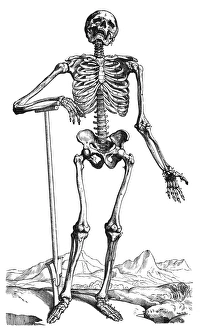
Background imageAnatomy Collection: Woodcut from the first book of Andreas Vesalius De Humani Corporis Fabrica

Woodcut from the first book of Andreas Vesalius De Humani Corporis Fabrica, published in 1543 at Basel
VESALIUS: SKELETAL SYSTEM. Woodcut from the first book of Andreas Vesalius De Humani Corporis Fabrica, published in 1543 at Basel

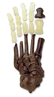
Background imageAnatomy Collection: Australopithecine or Homo habilis foot (OH8) cast

Australopithecine or Homo habilis foot (OH8) cast
Cast of a near complete foot (OH 8) from an Australopthecus or Homo habilis discovered at Olduvai Gorge, Tanzania by Louis Leakey in 1960. It dates back to 1.75 million years ago

Background imageAnatomy Collection: Muntiacus muntjak, Indian muntjac

Muntiacus muntjak, Indian muntjac
Zoological drawing 6 (1:6) Cervus plicatus. Pencil outline, lateral and front views, of head of Indian muntjac, made by Sydney Parkinson (1745-1771) during Captain Cooks first voyage of discovery

Background imageAnatomy Collection: Fire ant

Fire ant
Watercolour 405 by the Port Jackson Painter, entitled Mong, from the Watling Collection

Background imageAnatomy Collection: Phoenicurus phoenicurus, P. ochruros, Luscinia megarhynchos

Phoenicurus phoenicurus, P. ochruros, Luscinia megarhynchos
Plate 4 from Archibald Thorburns first edition of British Birds, Vol.1 (1915)

Background imageAnatomy Collection: Perdix perdix, grey partridge

Perdix perdix, grey partridge
Plate 13 from John Goulds The Birds of Great Britain, Vol. 4 (1873). Hand coloured lithograph

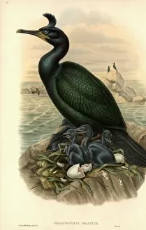
Background imageAnatomy Collection: Phalacrocorax aristotelis, European shag

Phalacrocorax aristotelis, European shag
Plate 53 from John Goulds The Birds of Great Britain, Vol. 5 (1873). Hand coloured lithograph

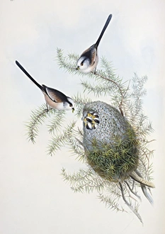
Background imageAnatomy Collection: Aegithalos caudatus, long-tailed tit

Aegithalos caudatus, long-tailed tit
Plate 28 from John Goulds The Birds of Great Britain, Vol. 2 (1873). Hand coloured lithograph

Background imageAnatomy Collection: Allosaurus cranium

Allosaurus cranium
A detail of the skull of Allosaurus, the Upper Jurassic carnivorous dinosaur that lived 153 to 135 million years ago. On display at the Natural History Museum, London

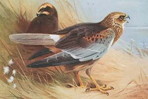
Background imageAnatomy Collection: Circus aeruginosus, marsh harrier

Circus aeruginosus, marsh harrier
Plate 73 from Archibald Thorburns second edtition of British Birds, Vol. 2 (1925)

Background imageAnatomy Collection: Booby

Booby
A pencil sketch from the Drawings Collection of Thomas Malie (1726-42)

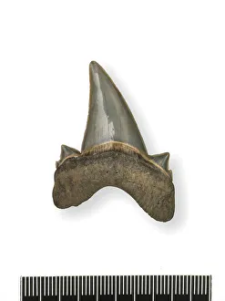
Background imageAnatomy Collection: Tonguestone (sharks tooth) with lateral denticles

Tonguestone (sharks tooth) with lateral denticles
Tooth of the extinct shark (Lamna obliqua Agassiz). Specimen orgininating from the London Clay (Eocene), Sheppey, Kent

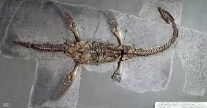
Background imageAnatomy Collection: Plesiosaurus hawkinsii

Plesiosaurus hawkinsii
A fossil specimen of the extinct marine reptile Plesiosaurus hawkinsii, on display at the Natural History Museum, London

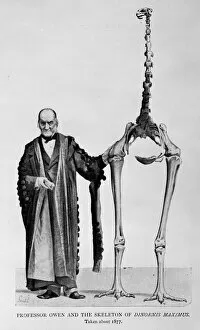
Background imageAnatomy Collection: Sir Richard Owen (1804-1892)

Sir Richard Owen (1804-1892)
Portrait of Sir Richard Owen, an English biologist, comparative anatomist and palaeontologist. Picture shows Owen and the skeleton of Dinornis maximus, c. 1877. From The Life of Owen (1894)

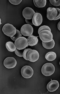
Background imageAnatomy Collection: Human red blood corpuscles

Human red blood corpuscles
Scanning electron microscope (SEM) of red blood cells showing their characteristic biconcave shape which increases the surface area for diffusion

Background imageAnatomy Collection: Central Hall

Central Hall
View of the Central Hall and Diplodocus replica skeleton, Gallery 10, Life Galleries at The Natural History Museum, London. The Museum was first opened to the public in 1881


All Professionally Made to Order for Quick Shipping

"Unveiling the Intricacies of Anatomy: From Sensory Homunculus to Motor Homunculus" Step into the fascinating world of anatomy, where every detail tells a story. Explore the depths of human and animal structures as we delve into this captivating subject. Let's begin with Leonardo da Vinci's masterpiece, "Head of a Bear. " Created in 1480 but discovered in 1945, this artwork showcases da Vinci's unparalleled ability to capture anatomical accuracy. The intricate details reveal his keen observation skills and deep understanding of form. Moving on to our furry friends, the greyhound takes center stage. Discovering the anatomy of these majestic creatures unveils their remarkable speed and agility. From their sleek skeleton to their muscular build, it is evident why they are renowned for their racing abilities. But what about us humans? Enter the sensory homunculus – a visual representation that depicts how our brain perceives different body parts based on sensitivity levels. This map reveals intriguing insights into how our senses are distributed throughout our bodies. X-ray images provide another dimension to understanding anatomy. Take a glimpse at normal knees through an X-ray lens – marvel at the complexity hidden beneath our skin while appreciating its delicate balance between strength and flexibility. The backbone is often considered one of nature's most ingenious designs. Our human backbone includes ribs and pelvis, providing stability while allowing movement – truly an architectural marvel worth exploring further. Venturing deeper within ourselves, let us explore the intricacies of the human brain from an inferior view. Witness its complex network of connections responsible for controlling various bodily functions - truly awe-inspiring. As we journey back in time, we encounter hominid crania – remnants that shed light on our evolutionary history. These ancient skulls offer glimpses into early forms such as Australopithecus afarensis (AL 288-1), famously known as Lucy - bridging gaps between past and present.