
Antibody Collection
"Unlocking the Power of Antibodies
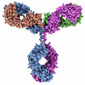
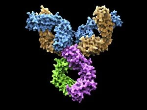
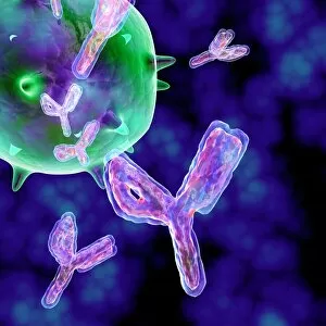
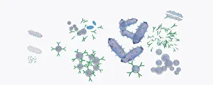
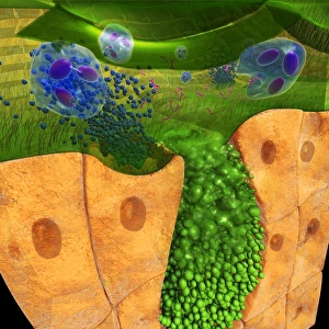
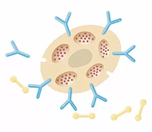
Illustration of response to infection after vaccination, involving microbe, antigens, antibody, plas


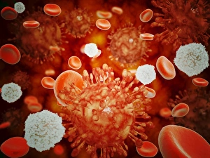
Digital cross section illustration of ciliate cell showing rhinovirus and antobodies in nasal cavity


All Professionally Made to Order for Quick Shipping
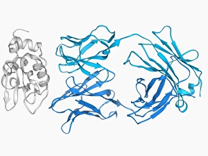
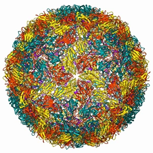
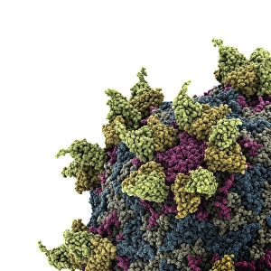
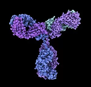
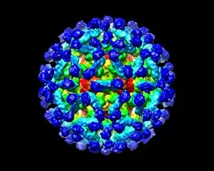
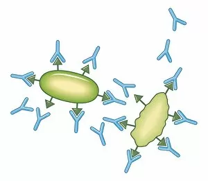
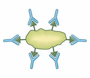
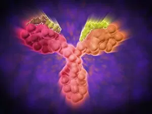
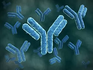
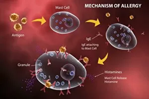
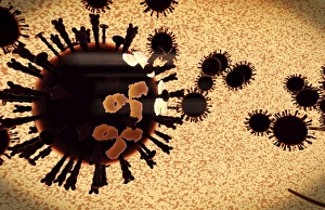
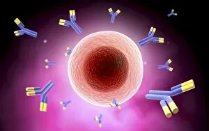
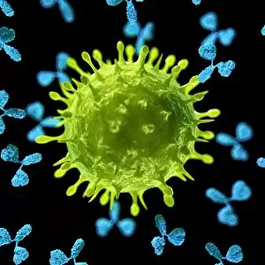
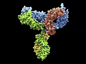
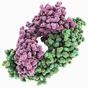
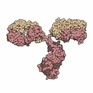
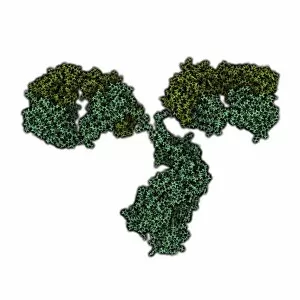
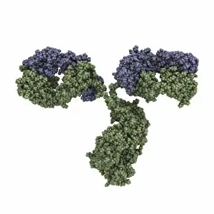
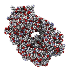
"Unlocking the Power of Antibodies: Exploring the Intricate World of Immunoglobulin G Molecules" Immerse yourself in the fascinating realm of antibodies with this captivating artwork showcasing the Immunoglobulin G antibody molecule. With its intricate structure and vital role in our immune system, these molecules are truly remarkable. Delve deeper into the world of immunology as you explore the Immunoglobulin G antibody molecule F007 / 9894. This detailed illustration highlights its unique features and emphasizes its significance in fighting off infections. Witness the incredible response to infection after vaccination through a stunning depiction involving microbes, antigens, antibodies, and plasma cells. This vivid artwork captures how our bodies mount a defense against harmful invaders, thanks to these powerful antibodies. Marvel at the complexity and versatility of Immunoglobulin G antibody molecules as they appear multiple times throughout this collection. Their ability to recognize specific pathogens is crucial for our immune system's effectiveness in combating diseases. Take a closer look at Rhinovirus and antibody molecular models C015 / 7139 as they interact within our bodies. These illustrations provide valuable insights into how antibodies neutralize viruses like rhinovirus, offering hope for future treatments or vaccines. Discover unexpected connections between Immunoglobulin G antibodies and egg whites (F006 / 9682). Unraveling such relationships sheds light on potential allergenic responses or even new avenues for research. Explore another intriguing aspect with Foot-and-mouth disease virus (F006 / 9556) where immunoglobulins play an essential role in diagnosing and treating this highly contagious viral infection that affects livestock worldwide. Continue your journey by examining Rhinovirus alongside an antibody molecular model C015 / 7138. Witness firsthand how these tiny proteins can bind to specific regions on viruses, preventing them from infecting healthy cells—an extraordinary defense mechanism. Conclude your exploration with yet another glimpse into the complex structure of an Immunoglobulin G antibody molecule C016 / 4462.