
Auditory Sense Collection
"Unveiling the Intricacies of Auditory Sense: Exploring the Inner Ear's Marvels" Delve into the captivating world as we unravel the hidden wonders within our inner ear


All Professionally Made to Order for Quick Shipping
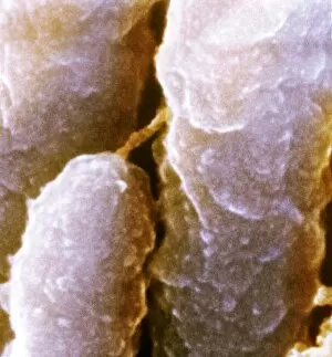
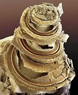
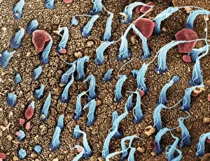
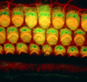
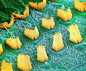
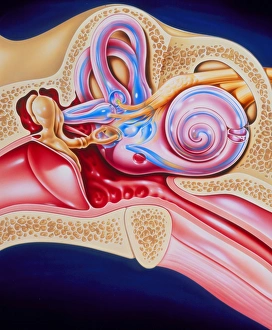
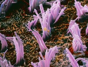
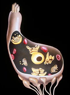
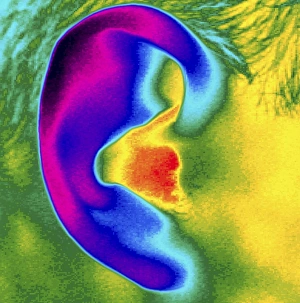
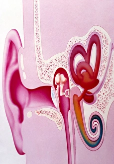
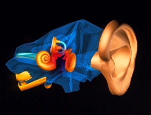
"Unveiling the Intricacies of Auditory Sense: Exploring the Inner Ear's Marvels" Delve into the captivating world as we unravel the hidden wonders within our inner ear. Through scanning electron microscopy (SEM), we can witness the intricate network of inner ear hairs, resembling delicate filaments that play a crucial role in our hearing. The Organ of Corti takes center stage, nestled within the depths of our inner ear. SEM imagery reveals its remarkable structure, showcasing rows upon rows of sensory hair cells meticulously arranged to capture sound vibrations and transform them into electrical signals for our brain to interpret. Embark on a visual journey through inner ear anatomy captured by C018 / 6400 SEM image – an awe-inspiring display unveiling the labyrinthine complexity that enables us to perceive sound. Witness how each component harmoniously contributes to this extraordinary system; from C018 / 6379 displaying cochlear ducts and spiral ganglion cells, to C018 / 6393 highlighting vestibular organs responsible for balance and spatial orientation. Marvel at C018 / 6387 capturing cochlear nerve fibers intertwining with blood vessels, illustrating their vital role in transmitting auditory information. Explore further with C018 / 6397 revealing detailed views of cochlea cross-sections, providing insight into its unique architecture. Witnessing these microscopic wonders doesn't end here - immerse yourself in C018 / 6402 SEM image showcasing vibrant inner ear hair cells. These specialized structures amplify sound waves and transmit them towards sensory cells depicted beautifully in another stunning SEM image. Even beyond these mesmerizing visuals lies yet another fascinating aspect: behold an up-close view of eardrum captured by SEM imaging techniques. Discover its intricate patterns and textures that enable it to vibrate when struck by sound waves – a gateway for auditory sensations. Intriguingly complex yet exquisitely designed, our auditory sense is truly a marvel of nature.