Fraud Blocker Skip to main content

Biochemical Collection (page 9)

"Unveiling the Intricacies of Biochemical World: From Anaesthetic Inhibiting Ion Channels to DNA Discoveries" Delving into the depths wonders

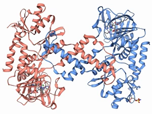
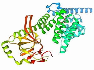
Background imageBiochemical Collection: Human lysine-specific demethylase F006 / 9747

Human lysine-specific demethylase F006 / 9747
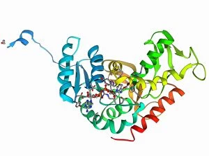
Human lysine-specific demethylase, molecular model. This enzyme removes methyl groups from lysine residues in histones (proteins that package DNA)

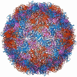
Background imageBiochemical Collection: Rhinovirus capsid, molecular model F006 / 9737

Rhinovirus capsid, molecular model F006 / 9737
Rhinovirus capsid, molecular model. This is human rhinovirus. The rhinovirus infects the upper respiratory tract and is the cause of the common cold. It is spread by coughs and sneezes

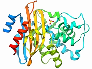
Background imageBiochemical Collection: Beta-lactamase molecule F006 / 9746

Beta-lactamase molecule F006 / 9746
Beta-lactamase enzyme, molecular model. This bacterial protein provides resistance to beta-lactam antibiotics, which include penicillin

Background imageBiochemical Collection: Human muscle aldolase, molecular model F006 / 9742

Human muscle aldolase, molecular model F006 / 9742
Human muscle aldolase. Molecular model of the enzyme human muscle aldolase complexed with its substrate fructose 1, 6-bisphosphate

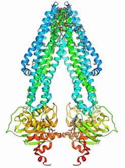
Background imageBiochemical Collection: ATP-binding cassette transporter F006 / 9743

ATP-binding cassette transporter F006 / 9743
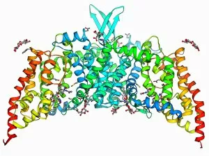
ATP-binding cassette transporter. Molecular model of the human mitochondrial ATP-binding cassette transporter ABCB10. This protein is found on the inner membrane of mitochondria

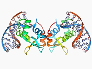
Background imageBiochemical Collection: Transcription factor bound to DNA F006 / 9744

Transcription factor bound to DNA F006 / 9744
Transcription factor bound to DNA. Molecular model of the human ETS translocation variant 1 (etv1) bound to a molecule of DNA (deoxyribonucleic acid)

Background imageBiochemical Collection: Beta-lactamase-like protein 2 molecule F006 / 9741

Beta-lactamase-like protein 2 molecule F006 / 9741
Beta-lactamase-like protein 2, molecular model

Background imageBiochemical Collection: Stress-responsive activator protein F006 / 9740

Stress-responsive activator protein F006 / 9740
Stress-responsive activator protein. Molecular model of the stress-responsive activator of p300 (strap) protein. This protein is activated when certain types of DNA (deoxyribonucleic acid)

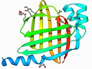
Background imageBiochemical Collection: Fatty acid binding protein 9 molecule F006 / 9739

Fatty acid binding protein 9 molecule F006 / 9739
Fatty acid binding protein 9. Molecular model of human testis-specific fatty acid binding protein 9

Background imageBiochemical Collection: Ebola virus glycoprotein and antibody F006 / 9738

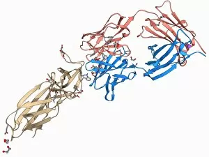
Ebola virus glycoprotein and antibody F006 / 9738
Ebola virus glycoprotein and antibody. Molecular model of a surface glycoprotein from the Ebola virus (EBOV) bound to an antibody

Background imageBiochemical Collection: Urea channel molecule F006 / 9735

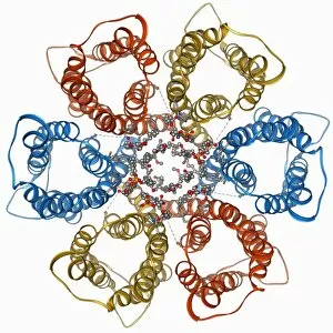
Urea channel molecule F006 / 9735
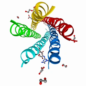
Urea channel. Molecular model of the proton-gated urea channel from the bacterium Helicobacter pylori. This channel opens in acidic environments, allowing the entry of urea into the cell

Background imageBiochemical Collection: Leptin receptor molecule F006 / 9736

Leptin receptor molecule F006 / 9736
Leptin receptor. Molecular model of a leptin receptor complexed with an antibody. Leptin is a hormone produced by adipose (fat) tissue

Background imageBiochemical Collection: DNA clamp complexed with DNA molecule F006 / 9732

DNA clamp complexed with DNA molecule F006 / 9732
DNA clamp complexed with DNA molecule. Molecular model showing a sliding DNA (deoxyribonucleic acid) clamp (ring) complexed with a molecule of DNA (blue and red)

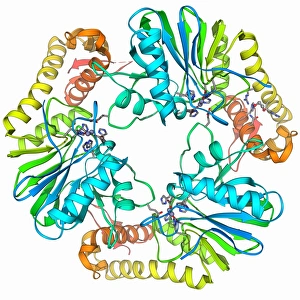
Background imageBiochemical Collection: GMP synthetase enzyme F006 / 9734

GMP synthetase enzyme F006 / 9734
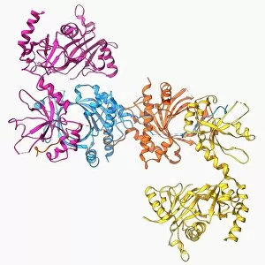
GMP synthetase enzyme, molecular model. This enzyme, guanine monophosphate synthetase, catalyses the reaction that converts xanthosine monophosphate to guanosine monophosphate

Background imageBiochemical Collection: Single stranded DNA-binding protein F006 / 9733

Single stranded DNA-binding protein F006 / 9733
Single stranded DNA-binding protein (SSBP). Molecular model of a protein that binds to the single stranded DNA (deoxyribonucleic acid) in human mitochondria

Background imageBiochemical Collection: Sir3 gene silencer acting on DNA F006 / 9730

Sir3 gene silencer acting on DNA F006 / 9730
Sir3 gene silencer acting on DNA, molecular model. Sir3 (light blue) is acting on a circular strand of DNA (deoxyribonucleic acid, pink)

Background imageBiochemical Collection: Parathion hydrolase enzyme F006 / 9731

Parathion hydrolase enzyme F006 / 9731
Parathion hydrolase, molecular model. This enzyme hydrolyses bonds in organophosphates, which include pesticides and the nerve gas sarin

Background imageBiochemical Collection: Tumour suppressor protein with DNA F006 / 9729

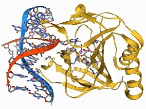
Tumour suppressor protein with DNA F006 / 9729
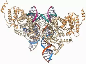
Tumour suppressor protein. Molecular model of the tumour suppressor protein p53 (beige) bound to a molecule of DNA (deoxyribonucleic acid, red and blue)

Background imageBiochemical Collection: Iron-regulatory protein bound to RNA F006 / 9727

Iron-regulatory protein bound to RNA F006 / 9727
Iron-regulatory protein bound to RNA, molecular model. Iron regulatory protein 1 (IRP1, purple) bound to a short strand of RNA (ribonucleic acid, red) that includes iron-responsive elements (IREs)

Background imageBiochemical Collection: DNA repair enzyme, molecular model F006 / 9726

DNA repair enzyme, molecular model F006 / 9726
DNA repair enzyme. Molecular model of the DNA (deoxyribonucleic acid) repair enzyme alpha-ketoglutarate-dependent dioxygenase bound to a molecule of DNA (red and blue)

Background imageBiochemical Collection: Oxidoreductase enzyme complex F006 / 9725

Oxidoreductase enzyme complex F006 / 9725
Oxidoreductase enzyme complex, molecular model. This is the membrane-bound domain formed from of a complex of NADH-quinone oxidoreductase subunits

Background imageBiochemical Collection: Saccharide transport protein F006 / 9722

Saccharide transport protein F006 / 9722
Saccharide transport protein. Molecular model of the phosphorylation-coupled saccharide transporter EIIC from the bacterium Bacillus cereus. EIIC is an integral membrane protein

Background imageBiochemical Collection: Aldo-keto reductase enzyme and ibuprofen F006 / 9724

Aldo-keto reductase enzyme and ibuprofen F006 / 9724
Aldo-keto reductase enzyme and ibuprofen. Molecular model of the enzyme aldo-keto reductase family 1 member C3 (AKR1C3) bound to a molecule of the anti-inflammatory drug ibuprofen

Background imageBiochemical Collection: SMAD4 protein domain bound to DNA F006 / 9723

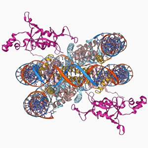
SMAD4 protein domain bound to DNA F006 / 9723
SMAD4 protein domain bound to DNA, molecular model. This strand of DNA (deoxyribonucleic acid, red and blue) is surrounded by MH1 domains of the SMAD4 (Mothers against decapentaplegic homolog 4)

Background imageBiochemical Collection: Transcription activator and DNA F006 / 9721

Transcription activator and DNA F006 / 9721
Transcription activator and DNA. Molecular model of the transcriptional activator of the multidrug efflux transporter BmrR bound to a molecule of DNA (deoxyribonucleic acid, red and blue)

Background imageBiochemical Collection: Methyltransferase complexed with DNA F006 / 9711

Methyltransferase complexed with DNA F006 / 9711
Methyltransferase complexed with DNA, molecular model. The strand of DNA (deoxyribonucleic acid, red and blue) is enclosed by DNA methyltransferase 1 (DNMT-1, beige)

Background imageBiochemical Collection: Pepsinogen molecule F006 / 9710

Pepsinogen molecule F006 / 9710
Pepsinogen. Molecular model of pepsinogen, the inactive precursor to the digestive enzyme pepsin. Pepsion, which is released by the stomach digests proteins

Background imageBiochemical Collection: Glycosylation enzyme molecule F006 / 9708

Glycosylation enzyme molecule F006 / 9708
Glycosylation enzyme. Molecular model of the enzyme N-acetylglucosamine (GlcNAc) transferase. This intracellular enzyme adds N-acetylglucosamine molecules to target proteins

Background imageBiochemical Collection: Retroviral intasome molecule F006 / 9705

Retroviral intasome molecule F006 / 9705
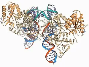
Retroviral intasome molecule. Molecular model of an intasome from a retrovirus complexed with host cell DNA (deoxyribonucleic acid)

Background imageBiochemical Collection: Retroviral intasome molecule F006 / 9706

Retroviral intasome molecule F006 / 9706
Retroviral intasome molecule. Molecular model of an intasome from a retrovirus complexed with host cell DNA (deoxyribonucleic acid)

Background imageBiochemical Collection: Metarhodopsin molecule F006 / 9709

Metarhodopsin molecule F006 / 9709
Metarhodopsin II molecule, molecular model. This is one of the intermediary breakdown products formed when light falls on the photoreceptor pigment rhodopsin

Background imageBiochemical Collection: NpmA methyltransferase F006 / 9707

NpmA methyltransferase F006 / 9707
NpmA methyltransferase, molecular model. Methyltransferase enzymes act to add methyl groups to nucleic acids such as DNA (deoxyribonucleic acid), a process called DNA methylation

Background imageBiochemical Collection: Notch transcription complex F006 / 9703

Notch transcription complex F006 / 9703
Notch transcription complex. Molecular model of the notch transcription complex bound to the human Hes1 promoter on a strand of DNA (deoxyribonucleic acid, red and blue)

Background imageBiochemical Collection: Oxidoreductase enzyme complex F006 / 9700

Oxidoreductase enzyme complex F006 / 9700
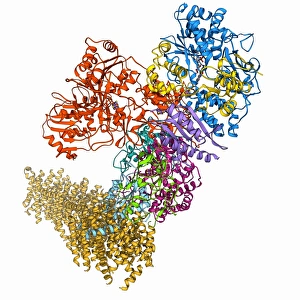
Oxidoreductase enzyme complex. Molecular model of a complex of NADH-quinone oxidoreductase subunits. The whole is termed respiratory complex I

Background imageBiochemical Collection: DNA repair enzyme, molecular model F006 / 9704

DNA repair enzyme, molecular model F006 / 9704
DNA repair enzyme. Molecular model of the DNA (deoxyribonucleic acid) repair enzyme dioxygenase bound to a molecule of DNA (red and blue)

Background imageBiochemical Collection: Lassa virus nucleocapsid protein F006 / 9702

Lassa virus nucleocapsid protein F006 / 9702
Lassa virus nucleocapsid protein, molecular model. This protein has essential roles in RNA (ribonucleic acid) synthesis and immune suppression

Background imageBiochemical Collection: Human rotavirus enterotoxin F006 / 9701

Human rotavirus enterotoxin F006 / 9701
Human rotavirus enterotoxin. Molecular model of the NSP4 (nonstructural protein 4) from the human rotavirus. This enterotoxin induces diarrhoea

Background imageBiochemical Collection: Isocitrate dehydrogenase kinase F006 / 9698

Isocitrate dehydrogenase kinase F006 / 9698
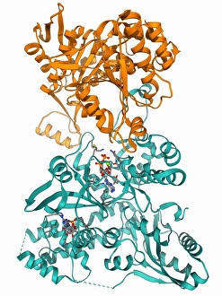
Isocitrate dehydrogenase kinase. Molecular model of isocitrate dehydrogenase kinase phosphatase (AceK) complexed with its substrate isocitrate dehydrogenase (ICDH)

Background imageBiochemical Collection: Ebola viral protein 35 and RNA F006 / 9697

Ebola viral protein 35 and RNA F006 / 9697
Ebola viral protein 35 and RNA. Molecular model of the Ebola viral protein 35 (VP35) bound to a molecule of double stranded RNA (ribonucleic acid)

Background imageBiochemical Collection: Cytochrome C, molecular model F006 / 9696

Cytochrome C, molecular model F006 / 9696
Cytochrome C, molecular model. Cytochrome molecules perform oxidation and reduction reactions for electron transport, a chain of reactions used to power cellular processes that require energy

Background imageBiochemical Collection: Lactate dehydrogenase enzyme molecule F006 / 9699

Lactate dehydrogenase enzyme molecule F006 / 9699
Lactate dehydrogenase enzyme, molecular model. This enzyme converts pyruvate to lactate in the final steps of glycolysis, and lactate to pyruvate during the lactic acid cycle

Background imageBiochemical Collection: mRNA capping apparatus F006 / 9694

mRNA capping apparatus F006 / 9694
mRNA capping apparatus. Molecular model of the Cet-1-Ceg1 mRNA capping apparatus

Background imageBiochemical Collection: Eye lens protein molecule F006 / 9695

Eye lens protein molecule F006 / 9695
Eye lens protein. Molecular model of alpha-crystallin, a protein found in the lens of the eye. The regular arrangement of the protein in the lens is thought to be responsible for its transparency

Background imageBiochemical Collection: Transcription repressor protein and DNA F006 / 9692

Transcription repressor protein and DNA F006 / 9692
Transcription repressor protein and DNA, molecular model. The repressor protein (green) is binding to a strand of DNA (deoxyribonucleic acid, pink and purple)

Background imageBiochemical Collection: Zinc transporter Yiip molecule F006 / 9691

Zinc transporter Yiip molecule F006 / 9691
Zinc transporter Yiip, molecular model. This transmembrane protein facilitates the movement of zinc ions

Background imageBiochemical Collection: Kinesin motor protein F006 / 9693

Kinesin motor protein F006 / 9693
Kinesin motor protein, molecular model. Kinesin motor proteins transport vesicles containing intracellular cargo around the cell along microtubules

Background imageBiochemical Collection: West Nile virus and antibodies F006 / 9689

West Nile virus and antibodies F006 / 9689
West Nile virus and antibodies. Molecular model of antibodies attached to the capsid of a west Nile virus. West Nile Virus is an arbovirus (arthropod-borne virus)

Background imageBiochemical Collection: Bluetongue virus capsid, molecular model F006 / 9687

Bluetongue virus capsid, molecular model F006 / 9687
Bluetongue virus capsid, molecular model. This virus, transmitted by midges, causes bluetongue disease in ruminants (commonly sheep, and less frequently cattle, goats and deer)


All Professionally Made to Order for Quick Shipping

"Unveiling the Intricacies of Biochemical World: From Anaesthetic Inhibiting Ion Channels to DNA Discoveries" Delving into the depths wonders, scientists have uncovered an anaesthetic that inhibits an ion channel (C015 / 6718), shedding light on new possibilities for pain management. The enigmatic double-stranded RNA molecule reveals its secrets, captivating researchers with its role in gene regulation and potential therapeutic applications. Peering into the intricate world of DNA transcription through a molecular model, scientists unravel the mysteries behind genetic information transfer and cellular function. Captured under a microscope's gaze, caffeine crystals dazzle with their vibrant beauty, reminding us of this ubiquitous stimulant's impact on our daily lives. The iconic DNA molecule stands tall as a symbol of life's blueprint, holding within it the key to our genetic heritage and evolutionary history. Immunoglobulin G antibody molecule emerges as a formidable defender against pathogens, showcasing nature's ingenious immune system at work. Through mesmerizing crystal formations seen under intense magnification, EDTA crystals reveal their significance in chelation therapy and metal ion sequestration processes. Oxytocin hormone crystals shimmer like precious gems when observed through polarized light microscopy (PLM C016 / 7196), highlighting its crucial role in social bonding and reproductive functions. Watson and Crick forever etched their names in scientific history by unravelling the structure of DNA; their groundbreaking discovery paved the way for countless advancements in genetics research. Celebrated author Isaac Asimov not only captivated readers with his science fiction tales but also left an indelible mark as a biochemist who popularized complex scientific concepts for all to comprehend and appreciate. Artistic renditions bring metabolic enzymes to life as they orchestrate vital chemical reactions within cells – true catalysts that drive life's intricate processes.