Fraud Blocker Skip to main content

Biochemical Collection (page 4)

"Unveiling the Intricacies of Biochemical World: From Anaesthetic Inhibiting Ion Channels to DNA Discoveries" Delving into the depths wonders

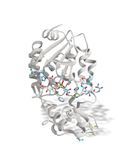
Background imageBiochemical Collection: SIRT3 molecule, artwork C017 / 3657

SIRT3 molecule, artwork C017 / 3657
SIRT3 molecule. Computer artwork showing the structure of a molecule of NAD-dependent deacetylase sirtuin-3, mitochondrial (SIRT3)

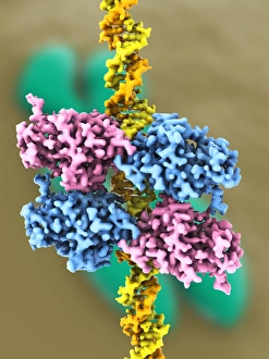
Background imageBiochemical Collection: Tumour suppressor protein and DNA C017 / 3646

Tumour suppressor protein and DNA C017 / 3646
Tumour suppressor protein and DNA. Computer artwork showing a molecule of the tumour suppressor protein p53 (blue and pink) bound to a molecule of DNA (deoxyribonucleic acid, yellow and orange)

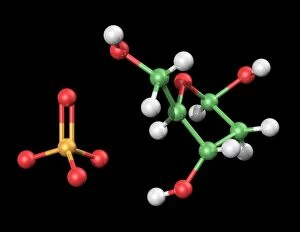
Background imageBiochemical Collection: DNA components, artwork C017 / 7350

DNA components, artwork C017 / 7350
DNA components. Computer artwork showing the structure of the two molecules that make up the backbone of DNA (deoxyribonucleic acid), phosphate (left) and deoxyribose (right)

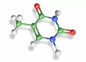
Background imageBiochemical Collection: Thymine molecule, artwork C017 / 7366

Thymine molecule, artwork C017 / 7366
Thymine molecule. Computer artwork showing the structure of a molecule of the nucleobase thymine. Atoms are colour-coded spheres: carbon (green), nitrogen (blue), oxygen (red), and hydrogen (white)

Background imageBiochemical Collection: Photosynthesis, artwork C017 / 0788

Photosynthesis, artwork C017 / 0788
Photosynthesis. Conceptual computer artwork of photosynthesis in a plant cell. Photosynthesis is the process by which most plants convert sunlight (coming from top left) into chemical energy

Background imageBiochemical Collection: Thymine molecule, artwork C017 / 7365

Thymine molecule, artwork C017 / 7365
Thymine molecule. Computer artwork showing the structure of a molecule of the nucleobase thymine. Atoms are colour-coded spheres: carbon (green), nitrogen (blue), oxygen (red), and hydrogen (white)

Background imageBiochemical Collection: Cytosine-guanine interaction, artwork C017 / 7216

Cytosine-guanine interaction, artwork C017 / 7216
Cytosine-guanine interaction. Computer artwork showing the structure of bound cytosine (left) and guanine molecules (right)

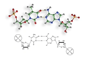
Background imageBiochemical Collection: Thymine-adenine interaction, artwork C017 / 7367

Thymine-adenine interaction, artwork C017 / 7367
Thymine-adenine interaction. Computer artwork showing the structure of bound thymine and adenine molecules. Atoms are shown as colour-coded spheres: carbon (green), hydrogen (white)

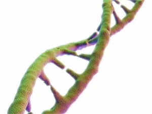
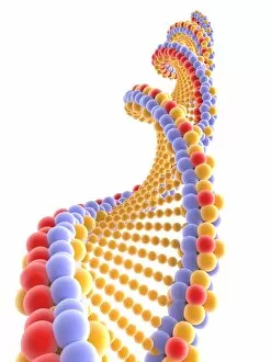
Background imageBiochemical Collection: DNA molecule, artwork F007 / 4200

DNA molecule, artwork F007 / 4200
DNA molecule, computer artwork

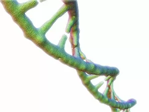
Background imageBiochemical Collection: DNA molecule, artwork F007 / 4196

DNA molecule, artwork F007 / 4196
DNA molecule, computer artwork

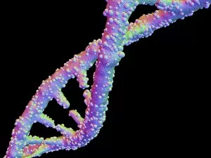
Background imageBiochemical Collection: DNA molecule, artwork F007 / 4203

DNA molecule, artwork F007 / 4203
DNA molecule, computer artwork

Background imageBiochemical Collection: DNA molecule, artwork F007 / 4207

DNA molecule, artwork F007 / 4207
DNA molecule, computer artwork

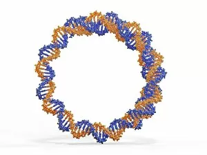
Background imageBiochemical Collection: Circular DNA molecule, artwork F006 / 7088

Circular DNA molecule, artwork F006 / 7088
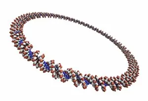
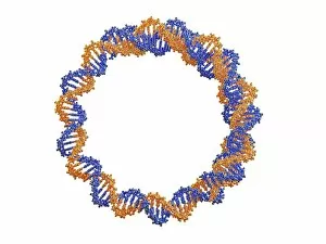
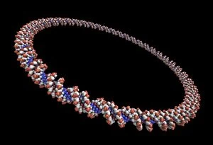
Circular DNA (deoxyribonucleic acid) molecule, computer artwork. Circular DNA has no ends, but consists of a ring structure

Background imageBiochemical Collection: Tablet computer showing a DNA molecule F006 / 6310

Tablet computer showing a DNA molecule F006 / 6310
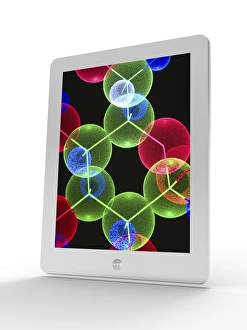
Tablet computer showing artwork of a DNA molecule

Background imageBiochemical Collection: Circular DNA molecule, space artwork F006 / 7089

Circular DNA molecule, space artwork F006 / 7089
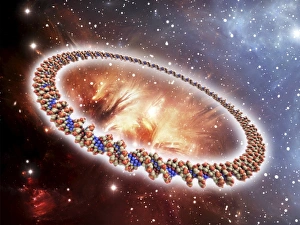
Circular DNA (deoxyribonucleic acid) molecule, computer artwork and space nebula artwork, depicting origin of life

Background imageBiochemical Collection: Tablet computer, insulin molecule F006 / 6311

Tablet computer, insulin molecule F006 / 6311
Tablet computer showing a part of the molecule of human insulin. A single insulin molecule is made up of two chains of amino acids, the A and B chains, which are held together by di-sulphide bridges

Background imageBiochemical Collection: Circular DNA molecule, artwork F006 / 7072

Circular DNA molecule, artwork F006 / 7072
Circular DNA (deoxyribonucleic acid) molecule, computer artwork. Circular DNA has no ends, but consists of a ring structure

Background imageBiochemical Collection: DNA molecule, artwork F006 / 3715

DNA molecule, artwork F006 / 3715
DNA molecule, computer artwork

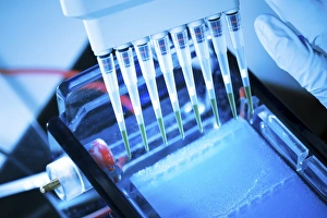
Background imageBiochemical Collection: Gel electrophoresis F006 / 7181

Gel electrophoresis F006 / 7181
Gel electrophoresis. Multipipette being used to place DNA (deoxyribonucleic acid) onto agarose gel

Background imageBiochemical Collection: DNA molecule, artwork F006 / 7147

DNA molecule, artwork F006 / 7147
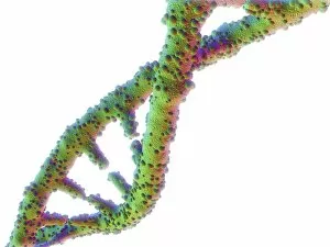
DNA (deoxyribonucleic acid) molecule, computer artwork

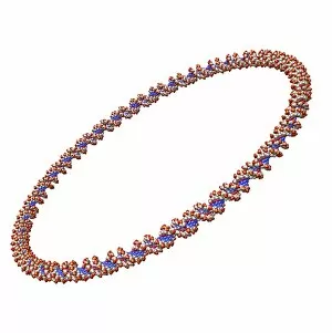
Background imageBiochemical Collection: Circular DNA molecule, artwork F006 / 7095

Circular DNA molecule, artwork F006 / 7095
Circular DNA (deoxyribonucleic acid) molecule, computer artwork. Circular DNA has no ends, but consists of a ring structure

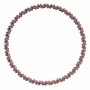
Background imageBiochemical Collection: Circular DNA molecule, artwork F006 / 7086

Circular DNA molecule, artwork F006 / 7086
Circular DNA (deoxyribonucleic acid) molecule, computer artwork. Circular DNA has no ends, but consists of a ring structure

Background imageBiochemical Collection: DNA molecule, artwork F006 / 3711

DNA molecule, artwork F006 / 3711
DNA molecule, computer artwork

Background imageBiochemical Collection: Circular DNA molecule, artwork F006 / 7083

Circular DNA molecule, artwork F006 / 7083
Circular DNA (deoxyribonucleic acid) molecule, computer artwork. Circular DNA has no ends, but consists of a ring structure

Background imageBiochemical Collection: Circular DNA molecule, space artwork F006 / 7077

Circular DNA molecule, space artwork F006 / 7077
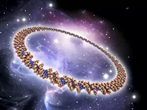
Circular DNA (deoxyribonucleic acid) molecule, computer artwork and space nebula artwork, depicting origin of life

Background imageBiochemical Collection: Circular DNA molecule, artwork F006 / 7084

Circular DNA molecule, artwork F006 / 7084
Circular DNA (deoxyribonucleic acid) molecule, computer artwork. Circular DNA has no ends, but consists of a ring structure

Background imageBiochemical Collection: Circular DNA molecule, space artwork F006 / 7087

Circular DNA molecule, space artwork F006 / 7087
Circular DNA (deoxyribonucleic acid) molecule, computer artwork and space nebula artwork, depicting origin of life

Background imageBiochemical Collection: DNA molecule, artwork F006 / 7127

DNA molecule, artwork F006 / 7127
DNA (deoxyribonucleic acid) molecule, computer artwork

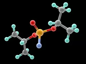
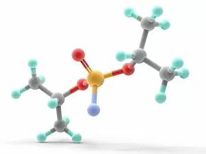
Background imageBiochemical Collection: Sarin nerve gas molecule F007 / 9936

Sarin nerve gas molecule F007 / 9936
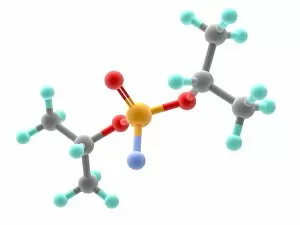
Sarin nerve gas, molecular model. Atoms are represented as spheres and are colour-coded: carbon (grey), hydrogen (green), oxygen (red), phosphorous (orange) and fluorine (blue)

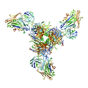
Background imageBiochemical Collection: Haemagglutinin viral surface protein F007 / 9932

Haemagglutinin viral surface protein F007 / 9932
Haemagglutinin viral surface protein. Molecular model of haemagglutinin, a surface protein from the influenza virus, complexed with a neutralising antibody

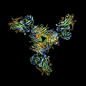
Background imageBiochemical Collection: Haemagglutinin viral surface protein F007 / 9931

Haemagglutinin viral surface protein F007 / 9931
Haemagglutinin viral surface protein. Molecular model of haemagglutinin, a surface protein from the influenza virus, complexed with a neutralising antibody

Background imageBiochemical Collection: Sarin nerve gas molecule F007 / 9933

Sarin nerve gas molecule F007 / 9933
Sarin nerve gas, molecular model. Atoms are represented as spheres and are colour-coded: carbon (grey), hydrogen (green), oxygen (red), phosphorous (orange) and fluorine (blue)

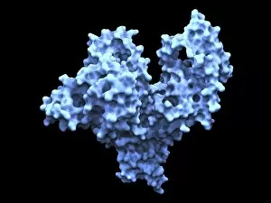
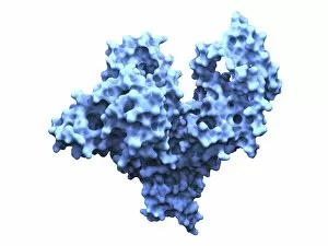
Background imageBiochemical Collection: Human serum albumin molecule F007 / 9904

Human serum albumin molecule F007 / 9904
Human serum albumin, molecular model. Albumin is the most abundant protein in human blood plasma. One of albumins functions is to transport fatty acids to the liver

Background imageBiochemical Collection: Human 80S ribosome F007 / 9902

Human 80S ribosome F007 / 9902
Ribosomal subunit. Computer model showing the structure of the RNA (ribonucleic acid) molecules in an 80S (large) ribosomal sub-unit. Ribosomes are composed of protein and RNA

Background imageBiochemical Collection: Human serum albumin molecule F007 / 9905

Human serum albumin molecule F007 / 9905
Human serum albumin, molecular model. Albumin is the most abundant protein in human blood plasma. One of albumins functions is to transport fatty acids to the liver

Background imageBiochemical Collection: Sarin nerve gas molecule F007 / 9938

Sarin nerve gas molecule F007 / 9938
Leucine, molecular model. Essential alpha-amino acid contained in eggs, soy protein, seaweed, turkey, chicken, lamb, cheese, and fish

Background imageBiochemical Collection: Human 80S ribosome F007 / 9898

Human 80S ribosome F007 / 9898
Ribosomal subunit. Computer model showing the structure of the RNA (ribonucleic acid) molecules in an 80S (large) ribosomal sub-unit. Ribosomes are composed of protein and RNA

Background imageBiochemical Collection: Sarin nerve gas molecule F007 / 9935

Sarin nerve gas molecule F007 / 9935
Sarin nerve gas, molecular model, The wire-frame map represents the electrostatic potential across the molecules surface. The atoms carbon, hydrogen, oxygen

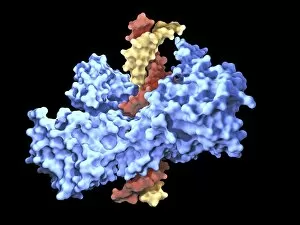
Background imageBiochemical Collection: Type I topoisomerase protein bound to DNA F007 / 9893

Type I topoisomerase protein bound to DNA F007 / 9893
Type I topoisomerase bound to DNA. Molecular model showing a type I topoisomerase molecule (blue) bound to a strand of DNA (deoxyribonucleic acid, yellow and red)

Background imageBiochemical Collection: DNA molecule, artwork F008 / 2034

DNA molecule, artwork F008 / 2034
DNA molecule, computer artwork

Background imageBiochemical Collection: DNA molecule, artwork F008 / 2036

DNA molecule, artwork F008 / 2036
DNA molecule, computer artwork

Background imageBiochemical Collection: DNA molecule, artwork F008 / 2040

DNA molecule, artwork F008 / 2040
DNA molecule, computer artwork

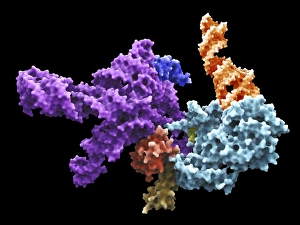
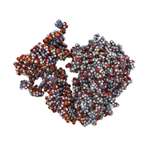
Background imageBiochemical Collection: Transfer RNA-synthetase complex molecule

Transfer RNA-synthetase complex molecule. Molecular model of a human tryptophanyl-tRNA synthetase molecule (red) complexed with a tRNA(Trp) molecule (blue)

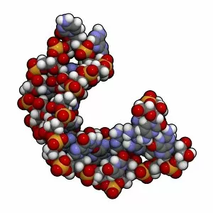
Background imageBiochemical Collection: MicroRNA precursor molecule

MicroRNA precursor molecule
MicroRNA (miRNA) precursor, molecular model. This miRNA (micro ribonucleic acid) precursor will be further processed into an even shorter mature miRNA oligonucleotide that can regulate the expression

Background imageBiochemical Collection: MicroRNA molecule

MicroRNA molecule
MicroRNA (miRNA), molecular model. This miRNA (micro ribonucleic acid) oligonucleotide regulates the expression of a target gene

Background imageBiochemical Collection: DNA nucleosome molecule

DNA nucleosome molecule
DNA nucleosome, molecular model. This is the fundamental repeating unit used to package DNA (deoxyribonucleic acid) inside cell nuclei

Background imageBiochemical Collection: tRNA molecule

tRNA molecule
Transfer RNA (tRNA), molecular model. tRNA (transfer ribonucleic acid) translates messenger RNA (mRNA) into a protein product. Each tRNA molecule carries a specific amino acid, in this case tryptophan

Background imageBiochemical Collection: Blood sample F005 / 0839

Blood sample F005 / 0839
MODEL RELEASED. Blood sample. Researcher extracting blood from a vial


All Professionally Made to Order for Quick Shipping

"Unveiling the Intricacies of Biochemical World: From Anaesthetic Inhibiting Ion Channels to DNA Discoveries" Delving into the depths wonders, scientists have uncovered an anaesthetic that inhibits an ion channel (C015 / 6718), shedding light on new possibilities for pain management. The enigmatic double-stranded RNA molecule reveals its secrets, captivating researchers with its role in gene regulation and potential therapeutic applications. Peering into the intricate world of DNA transcription through a molecular model, scientists unravel the mysteries behind genetic information transfer and cellular function. Captured under a microscope's gaze, caffeine crystals dazzle with their vibrant beauty, reminding us of this ubiquitous stimulant's impact on our daily lives. The iconic DNA molecule stands tall as a symbol of life's blueprint, holding within it the key to our genetic heritage and evolutionary history. Immunoglobulin G antibody molecule emerges as a formidable defender against pathogens, showcasing nature's ingenious immune system at work. Through mesmerizing crystal formations seen under intense magnification, EDTA crystals reveal their significance in chelation therapy and metal ion sequestration processes. Oxytocin hormone crystals shimmer like precious gems when observed through polarized light microscopy (PLM C016 / 7196), highlighting its crucial role in social bonding and reproductive functions. Watson and Crick forever etched their names in scientific history by unravelling the structure of DNA; their groundbreaking discovery paved the way for countless advancements in genetics research. Celebrated author Isaac Asimov not only captivated readers with his science fiction tales but also left an indelible mark as a biochemist who popularized complex scientific concepts for all to comprehend and appreciate. Artistic renditions bring metabolic enzymes to life as they orchestrate vital chemical reactions within cells – true catalysts that drive life's intricate processes.