Fraud Blocker Skip to main content

Biochemical Collection

"Unveiling the Intricacies of Biochemical World: From Anaesthetic Inhibiting Ion Channels to DNA Discoveries" Delving into the depths wonders

Background imageBiochemical Collection: DNA transcription, molecular model

DNA transcription, molecular model. Secondary structure of the enzyme RNA polymerase II synthesising a mRNA (messenger ribonucleic acid, lilac) strand from a DNA (deoxyribonucleic acid)

Background imageBiochemical Collection: Caffeine crystals, light micrograph

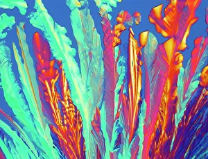
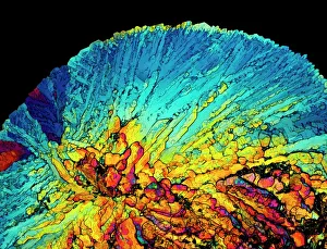
Caffeine crystals, light micrograph
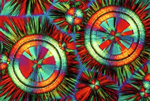
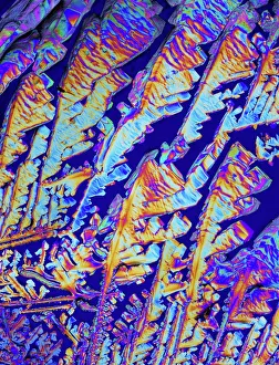
Caffeine crystals. Polarised light micrograph of crystals of caffeine (1, 3, 7-trimethylxanthine). Caffeine stimulates the central nervous system increasing alertness and deferring fatigue

Background imageBiochemical Collection: Anaesthetic inhibiting an ion channel C015 / 6718

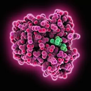
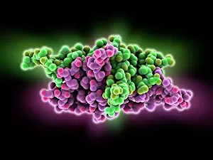
Anaesthetic inhibiting an ion channel C015 / 6718
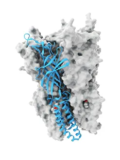
Anaesthetic inhibiting an ion channel. Computer model showing the structure of propofol anaesthetic drug molecules (spheres)

Background imageBiochemical Collection: Immunoglobulin G antibody molecule

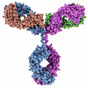
Immunoglobulin G antibody molecule. Computer model of the secondary structure of immunoglobulin G (IgG). This is the most abundant immunoglobulin and is found in all body fluids

Background imageBiochemical Collection: Double-stranded RNA molecule

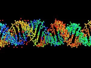
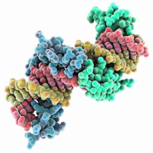
Double-stranded RNA molecule. Computer model of the structure of double-stranded RNA (ribonucleic acid). The majority of RNA in a cell is in the single-stranded form

Background imageBiochemical Collection: DNA molecule

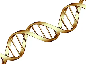
DNA molecule, computer artwork. DNA (deoxyribonucleic acid) is composed of two strands twisted into a double helix. Each strand consists of a sugar-phosphate backbone attached to nucleotide bases

Background imageBiochemical Collection: DNA molecule

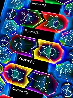
DNA molecule. Computer artwork of a molecule of DNA (deoxyribonucleic acid) with the chemical formulas of its components. DNA is composed of two sugar-phosphate backbones (blue)

Background imageBiochemical Collection: EDTA crystals, light micrograph

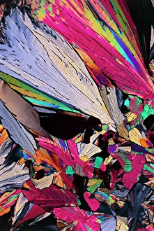
EDTA crystals, light micrograph
EDTA crystals. Polarised light micrograph of a section through ethylenediaminetetraacetic acid (EDTA) crystals. EDTA is used primarily for its effectiveness at binding metal ions (chelating)

Background imageBiochemical Collection: Watson and Crick, DNA discovers

Watson and Crick, DNA discovers
Watson and Crick. Caricature of the molecular biologists and discoverers of the structure of DNA James Watson (born 1928, left) and Francis Crick (1916-2004), with their model of a DNA molecule

Background imageBiochemical Collection: Isaac Asimov, US author and biochemist

Isaac Asimov, US author and biochemist
Isaac Asimov. Caricature of the Soviet-born American science fiction writer and biochemist Isaac Asimov (1920-1992). Asimov is best known for his science fiction novels and popular science books

Background imageBiochemical Collection: Metabolic enzyme, artwork

Metabolic enzyme, artwork
Metabolic enzyme. Computer artwork of aconitase (blue), in complex with ferritin messenger ribonucleic acid (mRNA, red). Aconitase is involved in the citric acid cycle but here it is performing a

Background imageBiochemical Collection: Secondary structure of proteins, artwork

Secondary structure of proteins, artwork
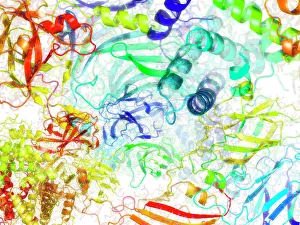
Secondary structure of proteins, computer artwork. The secondary structure is the shape taken by the strands of proteins, which are biological polymers of amino acids

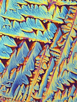
Background imageBiochemical Collection: Oxytocin hormone crystals, PLM C016 / 7196

Oxytocin hormone crystals, PLM C016 / 7196
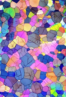
Oxytocin. Polarised light micrograph (PLM) of crystals of the female hormone oxytocin. In women this hormone is secreted naturally by the pituitary gland

Background imageBiochemical Collection: Cell membrane, artwork C013 / 7467

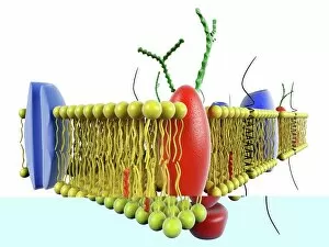
Cell membrane, artwork C013 / 7467
Computer artwork of a cutaway view of the human cell membrane. The cell Membrane is a complex part of the cell that controls what can get in and out of the cell

Background imageBiochemical Collection: Brain protein research

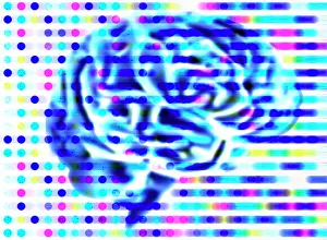
Brain protein research. Computer artwork of a brain and coloured dots from a protein microarray. Protein microarrays can be used to follow protein interactions

Background imageBiochemical Collection: Computer artwork of a beta DNA segment and spheres

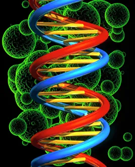
Computer artwork of a beta DNA segment and spheres
DNA molecule. Computer artwork of part of a strand of beta DNA (deoxyribonucleic acid) seen on a background of spheres. The spheres may represent bacteria

Background imageBiochemical Collection: Nucleotide base matrix

Nucleotide base matrix. Computer artwork depicting a matrix of nucleotide bases: adenine (A), cytosine (C), guanine (G) and thymine (T)

Background imageBiochemical Collection: DNA molecule, computer model

DNA molecule, computer model
DNA molecule. Computer artwork of the molecular structure of DNA (deoxyribonucleic acid). The DNA molecule is composed of two strands twisted into a double helix

Background imageBiochemical Collection: Nucleosome molecule

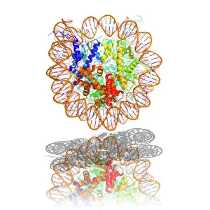
Nucleosome molecule, computer model. A nucleosome is a subunit of chromatin, the substance that forms chromosomes. It consists of a short length of DNA (deoxyribonucleic acid)

Background imageBiochemical Collection: DNA molecule, abstract image

DNA molecule, abstract image
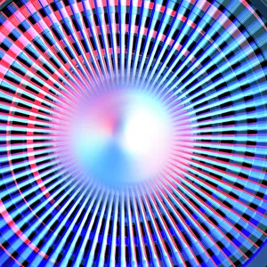
DNA molecule. Abstract computer artwork of a view along the inside of a molecule of DNA (deoxyribonucleic acid). DNA contains sections called genes that encode the bodys genetic information

Background imageBiochemical Collection: DNA nucleosome, molecular model

DNA nucleosome, molecular model
DNA nucleosome. Molecular model of a nucleosome, the fundamental repeating unit used to package DNA (deoxyribonucleic acid) inside cell nuclei

Background imageBiochemical Collection: Oxytocin crystals, light micrograph

Oxytocin crystals, light micrograph
Oxytocin. Polarised light micrograph of crystals of the female hormone oxytocin. In women this hormone is secreted naturally by the pituitary gland

Background imageBiochemical Collection: DNA molecule, artwork

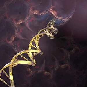
DNA molecule, artwork
DNA molecule. Computer artwork of a double stranded DNA (deoxyribonucleic acid) molecule amongst clouds of swirling gas. DNA is composed of two strands twisted into a double helix

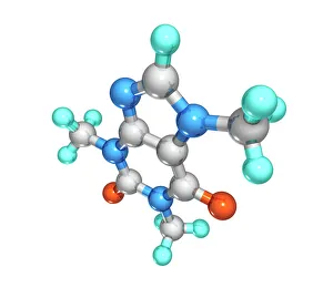
Background imageBiochemical Collection: Caffeine drug molecule

Caffeine drug molecule
Caffeine. Computer model of a molecule of the alkaloid, stimulant and legal drug caffeine. Caffeine is most often consumed in drinks like tea and coffee

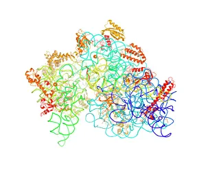
Background imageBiochemical Collection: Bacterial ribosome

Bacterial ribosome. Computer model showing the secondary structure of a 30S (small) ribosomal sub-unit from the bacteria Thermus thermophilus

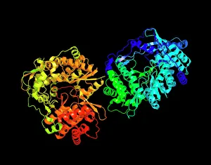
Background imageBiochemical Collection: HIV reverse transcription enzyme

HIV reverse transcription enzyme. Molecular models of the reverse transcriptase enzyme found in HIV (the human immunodeficiency virus)

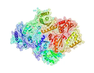
Background imageBiochemical Collection: Hepatitis C virus enzyme, molecular model

Hepatitis C virus enzyme, molecular model
Hepatitis C virus enzyme. Molecular model of a genetic enzyme from the Hepatitis C virus. This enzyme is called HC-J4 RNA polymerase

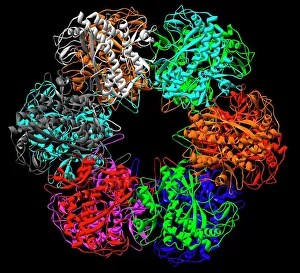
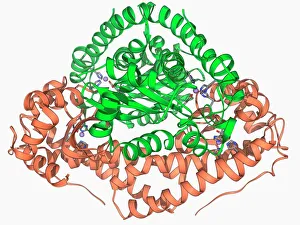
Background imageBiochemical Collection: Glutamine synthetase enzyme

Glutamine synthetase enzyme computer model. This is a ligase enzyme, which forms chemical bonds between molecules. The different colours show the different subunits that comprise the protein

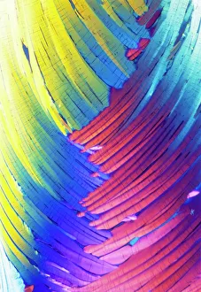
Background imageBiochemical Collection: Cortisol crystals, light micrograph

Cortisol crystals, light micrograph
Cortisol crystals, polarised light micrograph. Cortisol is a steroid hormone produced by the adrenal glands, which sit on top of the kidneys

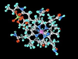
Background imageBiochemical Collection: Vitamin B12, molecular model

Vitamin B12, molecular model. Vitamin B12 (cyanocobalamin) is an essential nutrient that humans are unable to produce and need to obtain from their diet

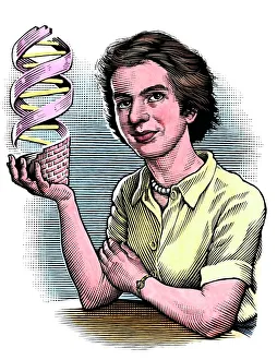
Background imageBiochemical Collection: Rosalind Franklin, British chemist

Rosalind Franklin, British chemist
Rosalind Franklin (1920-1958), British chemist and X-ray crystallographer, holding a model of DNA (deoxyribonucleic acid)

Background imageBiochemical Collection: DNA, illustration

DNA, illustration
DNA (deoxyribonucleic acid) molecule, computer illustration

Background imageBiochemical Collection: PLM of crystals of testosterone

PLM of crystals of testosterone
^BMale sex hormone.^b Polarised light micrograph of crystals of testosterone. Testosterone is the main human androgen, the class of steroid hormones responsible for the development of male

Background imageBiochemical Collection: RNA-editing enzyme, molecular model

RNA-editing enzyme, molecular model
RNA-editing enzyme. Molecular model of a left-handed, RNA double helix (Z-RNA, centre) bound by the Z alpha domain of the human RNA-editing enzyme ADAR1 (double-stranded RNA adenosine deaminase)

Background imageBiochemical Collection: Zinc fingers bound to a DNA strand

Zinc fingers bound to a DNA strand, molecular model. The double helix of DNA (deoxyribonucleic acid, red and yellow) is seen here with two Zif268 proteins (blue and green)

Background imageBiochemical Collection: Insulin crystals, light micrograph C017 / 8249

Insulin crystals, light micrograph C017 / 8249
Insulin. Polarised light micrograph (PLM) of crystals of the hormone insulin. The insulin molecule is made up of two chains of amino acids (A & B chains)

Background imageBiochemical Collection: Myoglobin molecule C015 / 5702

Myoglobin molecule C015 / 5702
Myoglobin molecule. Computer model showing the structure of a myoglobin molecule. Myoglobin is a protein found in muscle tissue

Background imageBiochemical Collection: DNA autoradiogram, artwork

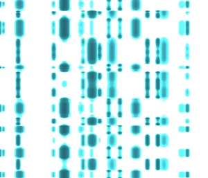
DNA autoradiogram, artwork. Autoradiograms show the order of nucleotide bases (basic building blocks) in a sample of DNA (deoxyribonucleic acid)

Background imageBiochemical Collection: Computer artwork of DNA replication

Computer artwork of DNA replication
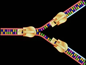
DNA replication. Computer artwork depicting DNA (deoxyribonucleic acid) replication. This segment of DNA is being " unzipped" to form a Y-shaped replication fork

Background imageBiochemical Collection: Capsaicin molecule

Capsaicin molecule
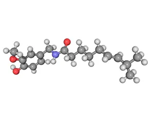
Capsaicin, molecular model. This chemical gives chilies their heat and causes a burning sensation when ingested. It is a secondary metabolite of the chili plant (Capsicum sp)

Background imageBiochemical Collection: Oxytocin neurotransmitter molecule

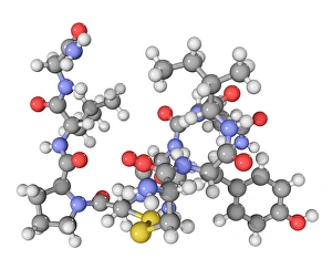
Oxytocin neurotransmitter molecule. Computer model showing the structure of the neurotransmitter and hormone Oxytocin. Atoms are colour-coded spheres (carbon: dark grey, hydrogen: light grey)

Background imageBiochemical Collection: Silver birch twig, SEM

Silver birch twig, SEM
Silver birch twig. Coloured scanning electron micrograph (SEM) of a section through a silver birch (Betula pendula) twig. At the centre of the twig (top centre left) is the pith

Background imageBiochemical Collection: Januvia diabetes drug molecule

Januvia diabetes drug molecule
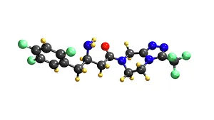
Januvia diabetes drug, molecular model. Januvia (sitagliptin) is a hypoglycaemic drug, one that reduces blood sugar levels

Background imageBiochemical Collection: Praziquantel parasite drug

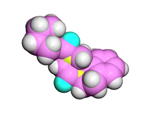
Praziquantel parasite drug. Computer model of a molecule of the drug praziquantel. Atoms are represented as spheres and are colour-coded; carbon (pink), hydrogen (white)

Background imageBiochemical Collection: Interferon molecule

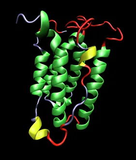
Interferon molecule. Computer model showing the secondary structure of a molecule of interferon. Interferons are proteins produced by white blood cells as part of the immune response to invading

Background imageBiochemical Collection: Oxytocin hormone crystals, LM C016 / 7195

Oxytocin hormone crystals, LM C016 / 7195
Oxytocin. Polarised light micrograph of crystals of the female hormone oxytocin. In women this hormone is secreted naturally by the pituitary gland

Background imageBiochemical Collection: Manganese superoxide dismutase enzyme F006 / 9423

Manganese superoxide dismutase enzyme F006 / 9423
Manganese superoxide dismutase enzyme, molecular model. This enzyme scavenges and decomposes the potentially toxic first reduction product, superoxide, of aerobic respiration

Background imageBiochemical Collection: SARS coronavirus protein

SARS coronavirus protein. Molecular model of the ORF-9b protein produced by the SARS (severe acute respiratory syndrome) coronavirus


All Professionally Made to Order for Quick Shipping

"Unveiling the Intricacies of Biochemical World: From Anaesthetic Inhibiting Ion Channels to DNA Discoveries" Delving into the depths wonders, scientists have uncovered an anaesthetic that inhibits an ion channel (C015 / 6718), shedding light on new possibilities for pain management. The enigmatic double-stranded RNA molecule reveals its secrets, captivating researchers with its role in gene regulation and potential therapeutic applications. Peering into the intricate world of DNA transcription through a molecular model, scientists unravel the mysteries behind genetic information transfer and cellular function. Captured under a microscope's gaze, caffeine crystals dazzle with their vibrant beauty, reminding us of this ubiquitous stimulant's impact on our daily lives. The iconic DNA molecule stands tall as a symbol of life's blueprint, holding within it the key to our genetic heritage and evolutionary history. Immunoglobulin G antibody molecule emerges as a formidable defender against pathogens, showcasing nature's ingenious immune system at work. Through mesmerizing crystal formations seen under intense magnification, EDTA crystals reveal their significance in chelation therapy and metal ion sequestration processes. Oxytocin hormone crystals shimmer like precious gems when observed through polarized light microscopy (PLM C016 / 7196), highlighting its crucial role in social bonding and reproductive functions. Watson and Crick forever etched their names in scientific history by unravelling the structure of DNA; their groundbreaking discovery paved the way for countless advancements in genetics research. Celebrated author Isaac Asimov not only captivated readers with his science fiction tales but also left an indelible mark as a biochemist who popularized complex scientific concepts for all to comprehend and appreciate. Artistic renditions bring metabolic enzymes to life as they orchestrate vital chemical reactions within cells – true catalysts that drive life's intricate processes.