Fraud Blocker Skip to main content

Cellular Collection (page 16)

"Unveiling the Intricacies of Cellular Life: A Journey Through Micrographs" Budding yeast cell: Witness the birth of new life as a budding yeast cell emerges

Background imageCellular Collection: Dandelion root, light micrograph

Dandelion root, light micrograph
Dandelion root. Light micrograph of a transverse section through a root of a dandelion (Taraxacum officinale). The root has undergone secondary thickening

Background imageCellular Collection: Vitreoscilla bacteria, TEM

Vitreoscilla bacteria, TEM
Vitreoscilla bacteria. Coloured transmission electron micrograph (TEM) of a section through several examples of Vitreoscilla sp. the gliding bacterium

Background imageCellular Collection: Spirogyra algae

Spirogyra algae
Spirogyra algal strand. Confocal light micrograph of a filament of Spirogyra sp. green algae. This alga forms slimy threads called " water-silk" or " mermaids tresses" in stagnant

Background imageCellular Collection: Woody stem section, SEM

Woody stem section, SEM
Woody stem section. Coloured scanning electron micrograph (SEM) of a section through a stem of a woody plant. At upper centre is the pith (orange), which consists of soft, spongy parenchyma cells

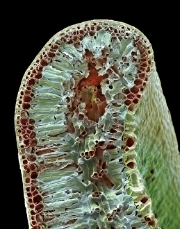
Background imageCellular Collection: Leaf tissue structure, SEM

Leaf tissue structure, SEM
Leaf tissue structure. Coloured scanning electron micrograph (SEM) of a section through the edge of a leaf from the Common Box (Buxus sempervirens)

Background imageCellular Collection: Leaf midrib, light micrograph

Leaf midrib, light micrograph
Leaf midrib. Light micrograph (LM) of a section through the midrib of a leaf from a monocotyledon plant. The midrib (midvein) is the continuation of a leafs stem along the centre of the leaf

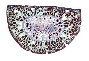
Background imageCellular Collection: Section through a pine needle, LM

Section through a pine needle, LM
Section through a pine needle. Light micrograph (LM) of a section through the needle (leaf) of a pine tree, Pinus sp.. The centre of the needle is occupied by two vascular bundles

Background imageCellular Collection: Leaf midrib, SEM

Leaf midrib, SEM
Leaf midrib. Coloured scanning electron micrograph (SEM) of a section through the midrib of a leaf from the Common Box (Buxus sempervirens)

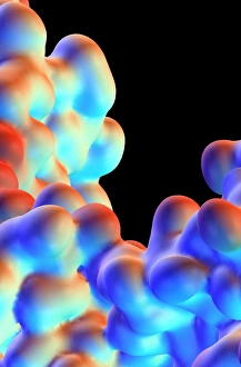
Background imageCellular Collection: ATPase muscle enzyme

ATPase muscle enzyme
Calcium pumping ATPase enzyme. Computer model of an electrostatic potential surface map of part of the ATPase enzyme that pumps calcium in and out of muscle cells and controls muscle contractions

Background imageCellular Collection: Mobile phone on charge, thermogram

Mobile phone on charge, thermogram. A thermogram shows the variation in temperature on the surface of an object, measured by the long-wave infrared radiation it emits


All Professionally Made to Order for Quick Shipping

"Unveiling the Intricacies of Cellular Life: A Journey Through Micrographs" Budding yeast cell: Witness the birth of new life as a budding yeast cell emerges, paving the way for future generations. Nerve and glial cells, light micrograph: Delve into the intricate network of nerve and glial cells that form the foundation of our nervous system. Dicotyledon plant stem, light micrograph: Explore the vibrant world within a dicotyledon plant stem, where cells work tirelessly to support growth and development. Rough endoplasmic reticulum, TEM: Peer into the fascinating realm of rough endoplasmic reticulum through a transmission electron microscope (TEM), revealing its role in protein synthesis. Dendritic cells, artwork: Marvel at an artistic representation showcasing dendritic cells' unique shape and function in immune responses. Cell structure: Uncover the hidden beauty within cellular structures as we unravel their complexity and significance in various biological processes. Mitosis, light micrograph: Observe nature's remarkable spectacle – mitosis – captured under a light microscope; witness how one cell divides into two with precision and grace. Cell division, fluorescent micrograph: Immerse yourself in a mesmerizing display of fluorescent colors capturing another facet of cell division's captivating dance. Dohle bodies in blood cell, micrograph: Discover peculiar "Dohle bodies" within blood cells through high-resolution microscopy—a clue to certain health conditions requiring further investigation. Acute promyelocytic leukemia, micrograph : Gain insight into acute promyelocytic leukemia by examining abnormal cellular characteristics observed under powerful magnification—an invaluable tool for diagnosis and treatment strategies Phagocytosis of fungal spores SEM : Witness nature's defense mechanism unfold as fungal spores are engulfed by cells in a captivating scanning electron microscope (SEM) image.