Fraud Blocker Skip to main content

Cellular Collection (page 7)

"Unveiling the Intricacies of Cellular Life: A Journey Through Micrographs" Budding yeast cell: Witness the birth of new life as a budding yeast cell emerges

Background imageCellular Collection: Potassium ion channel

Potassium ion channel. Computer artwork of a KcsA potassium ion (K+) channel (ribbons) embedded in a phospholipid (spheres) cell membrane (horizontal, centre)

Background imageCellular Collection: Voltage-gated potassium channel

Voltage-gated potassium channel. Computer model showing the molecular structure of a voltage-gated potassium (Kv) ion channel

Background imageCellular Collection: Endometrial hyperplasia, light micrograph C015 / 7104

Endometrial hyperplasia, light micrograph C015 / 7104
Endometrial hyperplasia. Light micrograph of a section through tissue from the endometrium, in a case of endometrial hyperplasia. The endometrium is the uterus lining

Background imageCellular Collection: Sir3 gene silencer acting on DNA C015 / 7062

Sir3 gene silencer acting on DNA C015 / 7062
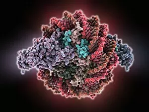
Sir3 gene silencer acting on DNA, molecular model. Sir3 (purple and grey) is acting on a circular strand of DNA (deoxyribonucleic acid, red)

Background imageCellular Collection: Sir3 gene silencer acting on DNA C015 / 7061

Sir3 gene silencer acting on DNA C015 / 7061
Sir3 gene silencer acting on DNA

Background imageCellular Collection: TFAM transcription factor bound to DNA C015 / 7060

TFAM transcription factor bound to DNA C015 / 7060
TFAM transcription factor bound to DNA, molecular model. Human mitochondrial transcription factor A (TFAM, pink) bound to a strand of DNA (deoxyribonucleic acid, orange and green)

Background imageCellular Collection: Peroxiredoxin 4 antioxidant enzyme C015 / 7022

Peroxiredoxin 4 antioxidant enzyme C015 / 7022
Peroxiredoxin 4 antioxidant enzyme, molecular model. This enzyme, also called peroxiredoxin IV (PrxIV), plays a catalytic role in cell metabolism on the endoplasmic reticulum

Background imageCellular Collection: Nucleus and endoplasmic reticulum C015 / 6797

Nucleus and endoplasmic reticulum C015 / 6797
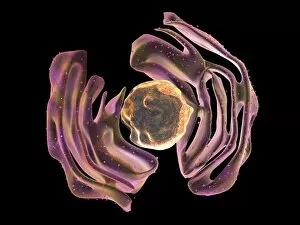
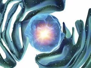
Computer artwork showing part of a human or eukaryotic cell. In the middle the nucleus which has a membrane with nuclear pores. Inside the nucleus is the DNA

Background imageCellular Collection: Animal cell organelles, artwork C015 / 6793

Animal cell organelles, artwork C015 / 6793
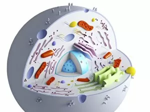
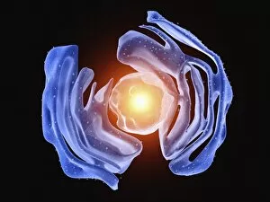
Animal cell organelles. Computer artwork showing the organelles in a eukaryotic cell. This is an animal cell. Structures include the nucleus (centre) which has a membrane with nuclear pores

Background imageCellular Collection: Animal cell organelles, artwork C015 / 6792

Animal cell organelles, artwork C015 / 6792
Animal cell organelles. Computer artwork showing the organelles in a eukaryotic cell. This is an animal cell. Structures include the nucleus (centre) which has a membrane with nuclear pores

Background imageCellular Collection: Nucleus and endoplasmic reticulum C015 / 6783

Nucleus and endoplasmic reticulum C015 / 6783
Computer artwork showing part of a human or eukaryotic cell. In the middle the nucleus which has a membrane with nuclear pores. Inside the nucleus is the DNA

Background imageCellular Collection: Nucleus and endoplasmic reticulum C015 / 6782

Nucleus and endoplasmic reticulum C015 / 6782
Computer artwork showing part of a human or eukaryotic cell. In the middle the nucleus which has a membrane with nuclear pores. Inside the nucleus is the DNA

Background imageCellular Collection: Nucleus and endoplasmic reticulum C015 / 6781

Nucleus and endoplasmic reticulum C015 / 6781
Computer artwork showing part of a human or eukaryotic cell. In the middle the nucleus which has a membrane with nuclear pores. Inside the nucleus is the DNA

Background imageCellular Collection: Animal cell organelles, artwork C015 / 6775

Animal cell organelles, artwork C015 / 6775
Animal cell organelles. Computer artwork showing the organelles in a eukaryotic cell. This is an animal cell. Structures include the nucleus (centre) which has a membrane with nuclear pores

Background imageCellular Collection: Animal cell organelles, artwork C015 / 6778

Animal cell organelles, artwork C015 / 6778
Animal cell organelles. Computer artwork showing the organelles in a eukaryotic cell. This is an animal cell. Structures include the nucleus (centre) which has a membrane with nuclear pores

Background imageCellular Collection: Animal cell organelles, artwork C015 / 6777

Animal cell organelles, artwork C015 / 6777
Animal cell organelles. Computer artwork showing the organelles in a eukaryotic cell. This is an animal cell. Structures include the nucleus (centre) which has a membrane with nuclear pores

Background imageCellular Collection: Nucleus and endoplasmic reticulum C015 / 6772

Nucleus and endoplasmic reticulum C015 / 6772
Computer artwork showing part of a human or eukaryotic cell. In the middle the nucleus which has a membrane with nuclear pores. Inside the nucleus is the DNA

Background imageCellular Collection: Nucleus and endoplasmic reticulum C015 / 6768

Nucleus and endoplasmic reticulum C015 / 6768
Computer artwork showing part of a human or eukaryotic cell. In the middle the nucleus which has a membrane with nuclear pores. Inside the nucleus is the DNA

Background imageCellular Collection: Nucleus and endoplasmic reticulum C015 / 6767

Nucleus and endoplasmic reticulum C015 / 6767
Computer artwork showing part of a human or eukaryotic cell. In the middle the nucleus which has a membrane with nuclear pores. Inside the nucleus is the DNA

Background imageCellular Collection: Animal cell organelles, artwork C016 / 0619

Animal cell organelles, artwork C016 / 0619
Computer artwork showing the organelles in a eukaryotic cell. This is an animal cell. Structures include the nucleus (centre, red) which has a membrane with nuclear pores

Background imageCellular Collection: Animal cell organelles, artwork C016 / 0621

Animal cell organelles, artwork C016 / 0621
Computer artwork showing the organelles in a eukaryotic cell. This is an animal cell. Structures include the nucleus (centre, red) which has a membrane with nuclear pores

Background imageCellular Collection: Animal cell organelles, artwork C016 / 0620

Animal cell organelles, artwork C016 / 0620
Computer artwork showing the organelles in a eukaryotic cell. This is an animal cell. Structures include the nucleus (centre, orange) which has a membrane with nuclear pores

Background imageCellular Collection: Animal cell organelles, artwork C016 / 0617

Animal cell organelles, artwork C016 / 0617
Computer artwork showing the organelles in a eukaryotic cell. This is an animal cell. Structures include the nucleus (centre, red) which has a membrane with nuclear pores

Background imageCellular Collection: Animal cell organelles, artwork C016 / 0618

Animal cell organelles, artwork C016 / 0618
Computer artwork showing the organelles in a eukaryotic cell. This is an animal cell. Structures include the nucleus (centre, orange) which has a membrane with nuclear pores

Background imageCellular Collection: Animal cell organelles, artwork C016 / 0615

Animal cell organelles, artwork C016 / 0615
Computer artwork showing the organelles in a eukaryotic cell. This is an animal cell. Structures include the nucleus (centre, orange) which has a membrane with nuclear pores

Background imageCellular Collection: Animal cell organelles, artwork C016 / 0616

Animal cell organelles, artwork C016 / 0616
Computer artwork showing the organelles in a eukaryotic cell. This is an animal cell. Structures include the nucleus (centre, orange) which has a membrane with nuclear pores

Background imageCellular Collection: Animal cell organelles, artwork C016 / 0611

Animal cell organelles, artwork C016 / 0611
Computer artwork showing the organelles in a eukaryotic cell. This is an animal cell. Structures include the nucleus (centre, red) which has a membrane with nuclear pores

Background imageCellular Collection: Animal cell organelles, artwork C016 / 0612

Animal cell organelles, artwork C016 / 0612
Computer artwork showing the organelles in a eukaryotic cell. This is an animal cell. Structures include the nucleus (centre, orange) which has a membrane with nuclear pores

Background imageCellular Collection: Animal cell organelles, artwork C016 / 0605

Animal cell organelles, artwork C016 / 0605
Computer artwork showing the organelles in a eukaryotic cell. This is an animal cell. Structures include the nucleus (centre, red) which has a membrane with nuclear pores

Background imageCellular Collection: Animal cell organelles, artwork C016 / 0610

Animal cell organelles, artwork C016 / 0610
Computer artwork showing the organelles in a eukaryotic cell. This is an animal cell. Structures include the nucleus (centre, orange) which has a membrane with nuclear pores

Background imageCellular Collection: Aquaporin membrane protein C015 / 5922

Aquaporin membrane protein C015 / 5922
Aquaporin membrane protein, molecular model. Aquaporins are membrane proteins that form channels (lower right) that help water molecules pass in and out of cells

Background imageCellular Collection: Animal cell organelles, artwork C016 / 0604

Animal cell organelles, artwork C016 / 0604
Computer artwork showing the organelles in a eukaryotic cell. This is an animal cell. Structures include the nucleus (centre, red) which has a membrane with nuclear pores

Background imageCellular Collection: Animal cell organelles, artwork C016 / 0606

Animal cell organelles, artwork C016 / 0606
Computer artwork showing the organelles in a eukaryotic cell. This is an animal cell. Structures include the nucleus (centre, orange) which has a membrane with nuclear pores

Background imageCellular Collection: Heat shock transcription factor and DNA C015 / 5558

Heat shock transcription factor and DNA C015 / 5558
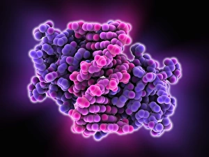
Heat shock transcription factor and DNA. Molecular model of the binding domain region (purple) of a heat shock protein transcription factor bound to DNA (pink, deoxyribonucleic acid)

Background imageCellular Collection: Oxidoreductase enzyme complex C015 / 6554

Oxidoreductase enzyme complex C015 / 6554
Oxidoreductase enzyme complex, molecular model. This is the membrane-bound domain formed from of a complex of NADH-quinone oxidoreductase subunits

Background imageCellular Collection: Oxidoreductase enzyme complex C015 / 6553

Oxidoreductase enzyme complex C015 / 6553
Oxidoreductase enzyme complex, molecular model. This is the membrane-bound domain formed from of a complex of NADH-quinone oxidoreductase subunits

Background imageCellular Collection: Vitamin B12 transport protein C015 / 5824

Vitamin B12 transport protein C015 / 5824
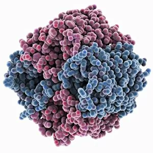
Vitamin B12 transport protein, molecular model. This transmembrane protein, known as BTUB, is from the Escherichia coli bacterium

Background imageCellular Collection: Vitamin B12 transport protein C015 / 5823

Vitamin B12 transport protein C015 / 5823
Vitamin B12 transport protein, molecular model. This transmembrane protein, known as BTUB, is from the Escherichia coli bacterium

Background imageCellular Collection: Customer in a mobile phone shop C018 / 2309

Customer in a mobile phone shop C018 / 2309
Shopping. A customer looking at mobile phones in a shop. Photographed in Moscow in 2012

Background imageCellular Collection: Caspase 6 enzyme C015 / 5700

Caspase 6 enzyme C015 / 5700
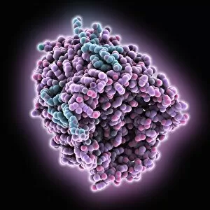
Capase 6 enzyme, molecular model. Caspases are proteases, enzymes that cleave proteins and play a role in apoptosis (programmed cell death)

Background imageCellular Collection: Caspase 6 enzyme C015 / 5699

Caspase 6 enzyme C015 / 5699
Capase 6 enzyme, molecular model. Caspases are proteases, enzymes that cleave proteins and play a role in apoptosis (programmed cell death)

Background imageCellular Collection: Cholera cytolysin cell toxin C015 / 6228

Cholera cytolysin cell toxin C015 / 6228
Cholera cytolysin cell toxin, molecular model. This is the Vibrio cholerae cytolysin (VCC) toxin from the bacterium that causes cholera

Background imageCellular Collection: Cholera cytolysin cell toxin C015 / 6229

Cholera cytolysin cell toxin C015 / 6229
Cholera cytolysin cell toxin, molecular model. This is the Vibrio cholerae cytolysin (VCC) toxin from the bacterium that causes cholera

Background imageCellular Collection: Sodium V-ATPase rotor ring C015 / 5379

Sodium V-ATPase rotor ring C015 / 5379
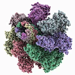
Sodium V-ATPase rotor ring, molecular model. V-ATPases (vacuolar-type ATPase) are evolutionarily ancient enzymes that helps transport ions across cell membranes

Background imageCellular Collection: Sodium V-ATPase rotor ring C015 / 5378

Sodium V-ATPase rotor ring C015 / 5378
Sodium V-ATPase rotor ring, molecular model. V-ATPases (vacuolar-type ATPase) are evolutionarily ancient enzymes that helps transport ions across cell membranes

Background imageCellular Collection: Chromatin remodelling factor and DNA C015 / 5156

Chromatin remodelling factor and DNA C015 / 5156
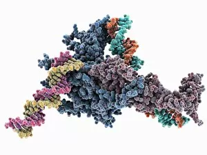
Chromatin remodelling factor and DNA, molecular model. The strands of DNA (deoxyribonucleic acid) are at left and right (both red and green). This chromatin remodelling factor (purple) is ISW1a

Background imageCellular Collection: Chromatin remodelling factor and DNA C015 / 5155

Chromatin remodelling factor and DNA C015 / 5155
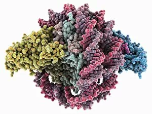
Chromatin remodelling factor and DNA, molecular model. The strands of DNA (deoxyribonucleic acid) are at left and right (pink-yellow and green-orange). This chromatin remodelling factor is ISW1a

Background imageCellular Collection: Importin protein complexed with enzyme C015 / 5149

Importin protein complexed with enzyme C015 / 5149
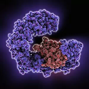
Importin protein complexed with enzyme, molecular model. Human importin13 (purple) complexed with SUMO-conjugating enzyme Ubc9 (red)


All Professionally Made to Order for Quick Shipping

"Unveiling the Intricacies of Cellular Life: A Journey Through Micrographs" Budding yeast cell: Witness the birth of new life as a budding yeast cell emerges, paving the way for future generations. Nerve and glial cells, light micrograph: Delve into the intricate network of nerve and glial cells that form the foundation of our nervous system. Dicotyledon plant stem, light micrograph: Explore the vibrant world within a dicotyledon plant stem, where cells work tirelessly to support growth and development. Rough endoplasmic reticulum, TEM: Peer into the fascinating realm of rough endoplasmic reticulum through a transmission electron microscope (TEM), revealing its role in protein synthesis. Dendritic cells, artwork: Marvel at an artistic representation showcasing dendritic cells' unique shape and function in immune responses. Cell structure: Uncover the hidden beauty within cellular structures as we unravel their complexity and significance in various biological processes. Mitosis, light micrograph: Observe nature's remarkable spectacle – mitosis – captured under a light microscope; witness how one cell divides into two with precision and grace. Cell division, fluorescent micrograph: Immerse yourself in a mesmerizing display of fluorescent colors capturing another facet of cell division's captivating dance. Dohle bodies in blood cell, micrograph: Discover peculiar "Dohle bodies" within blood cells through high-resolution microscopy—a clue to certain health conditions requiring further investigation. Acute promyelocytic leukemia, micrograph : Gain insight into acute promyelocytic leukemia by examining abnormal cellular characteristics observed under powerful magnification—an invaluable tool for diagnosis and treatment strategies Phagocytosis of fungal spores SEM : Witness nature's defense mechanism unfold as fungal spores are engulfed by cells in a captivating scanning electron microscope (SEM) image.