Fraud Blocker Skip to main content

Cellular Collection (page 2)

"Unveiling the Intricacies of Cellular Life: A Journey Through Micrographs" Budding yeast cell: Witness the birth of new life as a budding yeast cell emerges

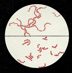
Background imageCellular Collection: Colony of Streptococcus pyogenes, 1906 (litho)

Colony of Streptococcus pyogenes, 1906 (litho)
3501894 Colony of Streptococcus pyogenes, 1906 (litho) by German School, (20th century); Private Collection; (add.info.: Bacteria constitute a large domain of prokaryotic microorganisms)

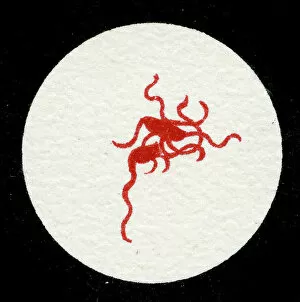
Background imageCellular Collection: Colony of Yersinia pestis, 1906 (litho)

Colony of Yersinia pestis, 1906 (litho)
3501917 Colony of Yersinia pestis, 1906 (litho) by German School, (20th century); Private Collection; (add.info.: Bacteria constitute a large domain of prokaryotic microorganisms)

Background imageCellular Collection: Colony of Salmonella Typhi showing Bacilli with flagella, 1906 (litho)

Colony of Salmonella Typhi showing Bacilli with flagella, 1906 (litho)
3501916 Colony of Salmonella Typhi showing Bacilli with flagella, 1906 (litho) by German School, (20th century); Private Collection; (add.info)

Background imageCellular Collection: Colony of Salmonella typhi, 1906 (litho)

Colony of Salmonella typhi, 1906 (litho)
3501915 Colony of Salmonella typhi, 1906 (litho) by German School, (20th century); Private Collection; (add.info.: Bacteria constitute a large domain of prokaryotic microorganisms)

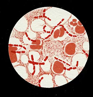
Background imageCellular Collection: Colony of Bacillus Anthracis in a human spleen, 1906 (litho)

Colony of Bacillus Anthracis in a human spleen, 1906 (litho)
3501905 Colony of Bacillus Anthracis in a human spleen, 1906 (litho) by German School, (20th century); Private Collection; (add.info)

Background imageCellular Collection: Spores of Bacillus Anthracis, 1906 (litho)

Spores of Bacillus Anthracis, 1906 (litho)
3501904 Spores of Bacillus Anthracis, 1906 (litho) by German School, (20th century); Private Collection; (add.info.: Bacteria constitute a large domain of prokaryotic microorganisms)

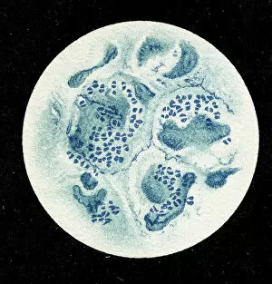
Background imageCellular Collection: Colony of Micrococcus Gonorrhoea found in the Urethra, 1906 (litho)

Colony of Micrococcus Gonorrhoea found in the Urethra, 1906 (litho)
3501900 Colony of Micrococcus Gonorrhoea found in the Urethra, 1906 (litho) by German School, (20th century); Private Collection; (add.info)

Background imageCellular Collection: Colony of Corynebacterium Diphtheriae, 1906 (litho)

Colony of Corynebacterium Diphtheriae, 1906 (litho)
3501907 Colony of Corynebacterium Diphtheriae, 1906 (litho) by German School, (20th century); Private Collection; (add.info.: Bacteria constitute a large domain of prokaryotic microorganisms)

Background imageCellular Collection: Colony of Bacterium mallei, 1906 (litho)

Colony of Bacterium mallei, 1906 (litho)
3501906 Colony of Bacterium mallei, 1906 (litho) by German School, (20th century); Private Collection; (add.info.: Bacteria constitute a large domain of prokaryotic microorganisms)

Background imageCellular Collection: Staphylococcus colony, 1905 (litho)

Staphylococcus colony, 1905 (litho)
3501895 Staphylococcus colony, 1905 (litho) by German School, (20th century); Private Collection; (add.info.: Bacteria constitute a large domain of prokaryotic microorganisms)

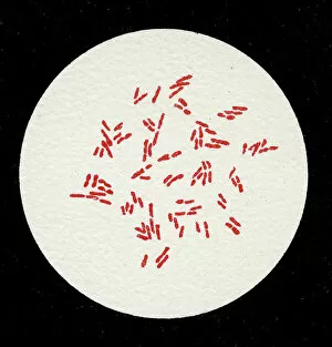
Background imageCellular Collection: Colony of Mycobacterium Tuberculosis, 1906 (litho)

Colony of Mycobacterium Tuberculosis, 1906 (litho)
3501911 Colony of Mycobacterium Tuberculosis, 1906 (litho) by German School, (20th century); Private Collection; (add.info.: Bacteria constitute a large domain of prokaryotic microorganisms)

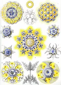
Background imageCellular Collection: Examples of Polycyttaria Kunstformen der Natur, 1899 (colour litho)

Examples of Polycyttaria Kunstformen der Natur, 1899 (colour litho)
746698 Examples of Polycyttaria Kunstformen der Natur, 1899 (colour litho) by Haeckel, Ernst (1824-1919) (after); Private Collection; (add.info.: Polycyttaria)

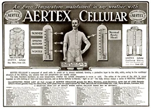
Background imageCellular Collection: Advert for Aertex Cellular Underwear and Shirts

Advert for Aertex Cellular Underwear and Shirts -- Clothed with Air. 1893

Background imageCellular Collection: The Cellular Clothing Co Ltd, Swindon

The Cellular Clothing Co Ltd, Swindon
Concerning Cotton - The Cellular Clothing Co Ltd - Swindon Factory. 1920

Background imageCellular Collection: The Cellular Clothing Co Ltd, Provost Street

The Cellular Clothing Co Ltd, Provost Street
Concerning Cotton - The Cellular Clothing Co Ltd - Provost Street Factory. 1920

Background imageCellular Collection: The Cellular Clothing Co Ltd, Fore Street, London

The Cellular Clothing Co Ltd, Fore Street, London
Concerning Cotton - The Cellular Clothing Co Ltd - Head Office, 72 Fore Street, London. 1920

Background imageCellular Collection: The Cellular Clothing Co Ltd, Basford

The Cellular Clothing Co Ltd, Basford
Concerning Cotton - The Cellular Clothing Co Ltd - Basford Factory. 1920

Background imageCellular Collection: The Cellular Clothing Co Ltd

The Cellular Clothing Co Ltd
Concerning Cotton - The Cellular Clothing Co Ltd. 1920

Background imageCellular Collection: Advert for Aertex mens underwear 1911

Advert for Aertex mens underwear 1911
Aertex Cellular clothing for men, for all seasons. Date: 1911

Background imageCellular Collection: Advert for Aertex men and womens underwear 1933

Advert for Aertex men and womens underwear 1933
Aertex underwear, little cells in the fabric that allow the body to breathe. Date: 1933

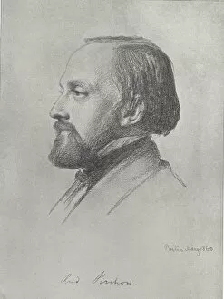
Background imageCellular Collection: Rudolf Ludwig Karl Virchow (litho)

Rudolf Ludwig Karl Virchow (litho)
815951 Rudolf Ludwig Karl Virchow (litho) by English School, (19th century); Private Collection; (add.info.: Rudolf Ludwig Karl Virchow (1821-1902).); Look and Learn / Elgar Collection

Background imageCellular Collection: Plan of the Mazas Prison and cells for prisoners, Boulevard Mazas, Paris (w / c on paper)

Plan of the Mazas Prison and cells for prisoners, Boulevard Mazas, Paris (w / c on paper)
XIR437861 Plan of the Mazas Prison and cells for prisoners, Boulevard Mazas, Paris (w/c on paper) by French School, (19th century); Musee de la Ville de Paris, Musee Carnavalet, Paris

Background imageCellular Collection: Botany (colour litho)

Botany (colour litho)
728983 Botany (colour litho) by English School, (19th century); Private Collection; (add.info.: Botany, with depictions of cellular cryptograms (1. fungus; 2. seaweed; 3)

Background imageCellular Collection: Boy Listening to Music

Boy Listening to Music

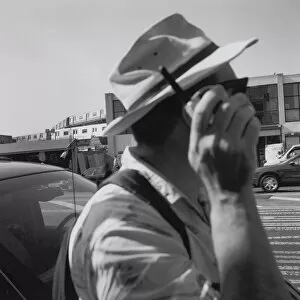
Background imageCellular Collection: Man with cell phone

Man with cell phone
adult, brunette, casual, cell phone, cellular, communication, computer, conversation, day, hand, head and shoulders, leisure, man, mobile, one person only, outdoor, side view, street, summer, talk

Background imageCellular Collection: Woman Taking a Selfie

Woman Taking a Selfie

Background imageCellular Collection: Advert for Aertex mens clothes with air 1895

Advert for Aertex mens clothes with air 1895
Cellular Aertex cloth shirts and underwear, which the air is enclosed and warmed by heat of the body. Date: 1895

Background imageCellular Collection: EyeUbiquitous_20122206

EyeUbiquitous_20122206
Transport, Cars, Electric, Charging point in car park

Background imageCellular Collection: EyeUbiquitous_20122211

EyeUbiquitous_20122211
Transport, Cars, Electric, Vehicle plugged into recharge point

Background imageCellular Collection: EyeUbiquitous_20122209

EyeUbiquitous_20122209
Transport, Cars, Electric, Recharge point

Background imageCellular Collection: EyeUbiquitous_20122208

EyeUbiquitous_20122208
Transport, Cars, Electric, Recharging point and sign

Background imageCellular Collection: EyeUbiquitous_20105670

EyeUbiquitous_20105670
Communications, Telephones, Mobile, Phone stuck between cushions of sofa

Background imageCellular Collection: EyeUbiquitous_20105669

EyeUbiquitous_20105669
Communications, Telephones, Mobile, Phone stuck between cushions of sofa

Background imageCellular Collection: EyeUbiquitous_20105671

EyeUbiquitous_20105671
Communications, Telephones, Mobile, Phone stuck between cushions of sofa

Background imageCellular Collection: EyeUbiquitous_20105668

EyeUbiquitous_20105668
Communications, Telephones, Mobile, Phone stuck between cushions of sofa

Background imageCellular Collection: Communications, Telephone, Mobile

Communications, Telephone, Mobile, Man using Apple iPhone apps

Background imageCellular Collection: EyeUbiquitous_20108355

EyeUbiquitous_20108355
Peru, Chinchero, Young woman in traditional dress using a cell phone to take a picture of herself and other woman with a child

Background imageCellular Collection: EyeUbiquitous_20107347

EyeUbiquitous_20107347
Peru, Woman with baby in traditional dress

Background imageCellular Collection: EyeUbiquitous_20108349

EyeUbiquitous_20108349
Peru, Young woman using a Cell Phone

Background imageCellular Collection: EyeUbiquitous_20108339

EyeUbiquitous_20108339
Vietnam, Two Young Ladies taking a picture of them selves, with a camera phone

Background imageCellular Collection: Spain, Madrid, Tourists pose for photographs in front of the Palacio Real

Spain, Madrid, Tourists pose for photographs in front of the Palacio Real

Background imageCellular Collection: India, West Bengal, Kolkata, A worker at a food hotel in takes a break to use his iphone

India, West Bengal, Kolkata, A worker at a food hotel in takes a break to use his iphone

Background imageCellular Collection: EyeUbiquitous_20108335

EyeUbiquitous_20108335
Laos, man in small village near the Mekong, having his picture taken by man with mobile telephone

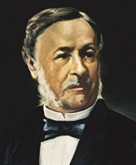
Background imageCellular Collection: SCHWANN, Theodor (1810 - 1882). German physiologist

SCHWANN, Theodor (1810 - 1882). German physiologist, anatomist and naturalist. He developed the cell theory. Oil

Background imageCellular Collection: TFAM transcription factor bound to DNA C015 / 7059

TFAM transcription factor bound to DNA C015 / 7059
TFAM transcription factor bound to DNA, molecular model. Human mitochondrial transcription factor A (TFAM, green) bound to a strand of DNA (deoxyribonucleic acid, blue and pink)

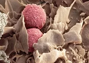
Background imageCellular Collection: Phagocytosis of fungus spores, SEM

Phagocytosis of fungus spores, SEM
Phagocytosis of fungus spores. Coloured scanning electron micrograph (SEM) of spores from the fungus Aspergillus fumigatus (red) being phagocytosed (engulfed and destroyed) by a dendritic cell (brown)

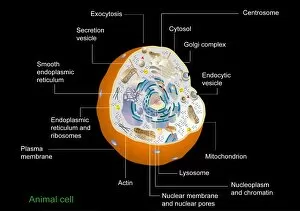
Background imageCellular Collection: Animal cell anatomy, diagram

Animal cell anatomy, diagram
Animal cell anatomy. Diagram showing the internal and external anatomy of an animal cell


All Professionally Made to Order for Quick Shipping

"Unveiling the Intricacies of Cellular Life: A Journey Through Micrographs" Budding yeast cell: Witness the birth of new life as a budding yeast cell emerges, paving the way for future generations. Nerve and glial cells, light micrograph: Delve into the intricate network of nerve and glial cells that form the foundation of our nervous system. Dicotyledon plant stem, light micrograph: Explore the vibrant world within a dicotyledon plant stem, where cells work tirelessly to support growth and development. Rough endoplasmic reticulum, TEM: Peer into the fascinating realm of rough endoplasmic reticulum through a transmission electron microscope (TEM), revealing its role in protein synthesis. Dendritic cells, artwork: Marvel at an artistic representation showcasing dendritic cells' unique shape and function in immune responses. Cell structure: Uncover the hidden beauty within cellular structures as we unravel their complexity and significance in various biological processes. Mitosis, light micrograph: Observe nature's remarkable spectacle – mitosis – captured under a light microscope; witness how one cell divides into two with precision and grace. Cell division, fluorescent micrograph: Immerse yourself in a mesmerizing display of fluorescent colors capturing another facet of cell division's captivating dance. Dohle bodies in blood cell, micrograph: Discover peculiar "Dohle bodies" within blood cells through high-resolution microscopy—a clue to certain health conditions requiring further investigation. Acute promyelocytic leukemia, micrograph : Gain insight into acute promyelocytic leukemia by examining abnormal cellular characteristics observed under powerful magnification—an invaluable tool for diagnosis and treatment strategies Phagocytosis of fungal spores SEM : Witness nature's defense mechanism unfold as fungal spores are engulfed by cells in a captivating scanning electron microscope (SEM) image.