
Confocal Collection
"Exploring the Intricacies of Cell Biology: A Journey through Confocal Micrographs" Step into the fascinating world microscopy


All Professionally Made to Order for Quick Shipping
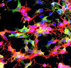
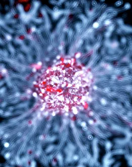
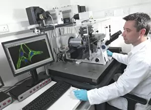
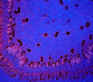
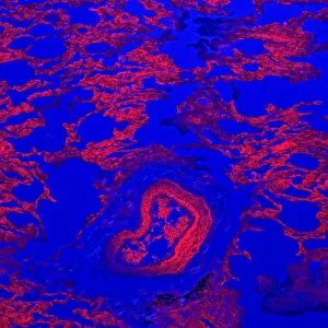
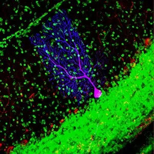
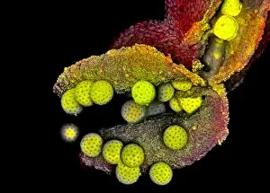
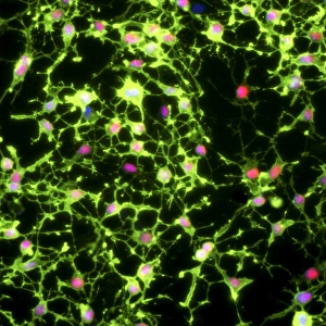
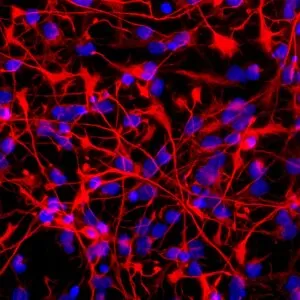
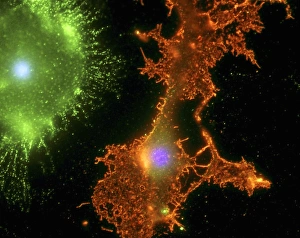
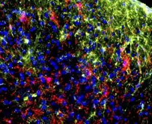
"Exploring the Intricacies of Cell Biology: A Journey through Confocal Micrographs" Step into the fascinating world microscopy, where scientists unravel the secrets hidden within cells. In this captivating collection of images, we delve into various aspects of cell biology and witness the beauty that lies beneath our skin. First, we encounter glial stem cell culture under the lens. The delicate network of cells reveals their potential to regenerate and support neurons in our nervous system. Next, neural stem cell culture showcases their remarkable ability to differentiate into different types of brain cells, offering hope for future therapies. Moving on to a bustling cell biology laboratory, researchers meticulously study cotton stems using confocal micrograph C014 / 4636. These high-resolution images provide insights into plant structures at a cellular level and aid in understanding their growth patterns. Shifting gears back to human anatomy, squamous epithelium takes center stage with confocal micrographs C014 / 4643 and C014 / 4642. These detailed snapshots allow us to appreciate the intricate layers that protect our organs from external harm. Venturing deeper inside our bodies, we explore tissues such as testis (C014 / 4617), lung (C014 / 4616), and kidney (C014 / 4611). Each image captures unique characteristics specific to these organs—testis revealing spermatogenesis intricacies; lung displaying its complex respiratory structure; and kidney showcasing its filtration capabilities. Zooming further into kidney tissue brings us face-to-face with its intricate blood vessels (C014 / 4609-4610). These mesmerizing networks highlight how essential they are for maintaining proper renal function by filtering waste products from our bloodstream. Confocal microscopy has revolutionized our understanding of cellular structures across various fields—from neuroscience to botany—and continues to push boundaries in scientific exploration. Through these breathtaking micrographs, we gain a glimpse into nature's complexity, reminding us of the wonders that lie within ourselves and the world around us.