Home > Popular Themes > Human Body
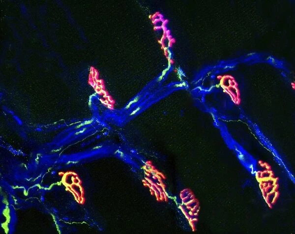
Neuromuscular synapse, light micrograph
![]()

Wall Art and Photo Gifts from Science Photo Library
Neuromuscular synapse, light micrograph
Neuromuscular junction. Fluorescent confocal light micrograph of the junction between a nerve cell and a muscle (not seen). The axon of the nerve cell (neuron) has been tagged with a blue dye. The axon ends at end plates, which form junctions called synapses with the muscle cells. The end plates have been dyed red here by tagging them with the snake venom alpha-bungarotoxin, which binds to them. When a nerve signal reaches the synapse, it causes the synaptic vesicles to rupture, releasing the neurotransmitter acetylcholine. The protein synaptophysin, found in the vesicles, is dyed green. Acetylcholine binds to receptors on the muscle cells, causing them to contract. Magnification: x120 when printed 10cm wide
Science Photo Library features Science and Medical images including photos and illustrations
Media ID 6449461
© THOMAS DEERINCK, NCMIR/SCIENCE PHOTO LIBRARY
Acetylcholine Axon Axons Confocal Dyed Dyes Fluorescent Light Micrograph Histological Histology Neuron Neurons Neurotransmitter Neurotransmitters Receptor Synapse Synapses Synaptic Vesicle Tagged Vesicles Bio Chemistry Light Microscope Nervous System Neurological Neurology
EDITORS COMMENTS
This print from Science Photo Library showcases the intricate beauty of a neuromuscular synapse. In this fluorescent confocal light micrograph, we witness the junction between a nerve cell and a muscle, although the muscle itself remains unseen. The nerve cell's axon has been meticulously tagged with a striking blue dye, drawing our attention to its vital role in transmitting signals. The end plates at the termination of the axon are dyed an intense red using snake venom alpha-bungarotoxin, which binds specifically to them. These end plates form synapses with the muscle cells, serving as crucial connection points for communication within our nervous system. Highlighted in vibrant green is synaptophysin, a protein found within synaptic vesicles that play an essential role in neurotransmission. When stimulated by a nerve signal, these vesicles rupture and release acetylcholine - an important neurotransmitter responsible for initiating muscular contractions. With magnification set at x120 when printed 10cm wide, this image allows us to marvel at the microscopic wonders hidden within our own bodies. It serves as a testament to both biology and neurology – showcasing how delicate interactions between neurons and muscles enable seamless movement and coordination. Science Photo Library continues to provide awe-inspiring visuals that bridge scientific knowledge with artistic appreciation.
MADE IN AUSTRALIA
Safe Shipping with 30 Day Money Back Guarantee
FREE PERSONALISATION*
We are proud to offer a range of customisation features including Personalised Captions, Color Filters and Picture Zoom Tools
SECURE PAYMENTS
We happily accept a wide range of payment options so you can pay for the things you need in the way that is most convenient for you
* Options may vary by product and licensing agreement. Zoomed Pictures can be adjusted in the Cart.

