
Confocal Light Micrograph Collection
"Exploring the Intricacies of Cerebellum Tissue


All Professionally Made to Order for Quick Shipping
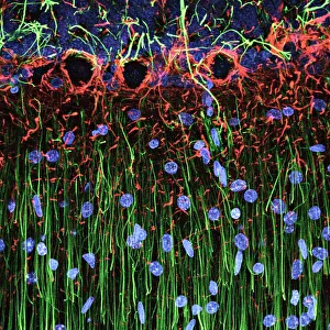
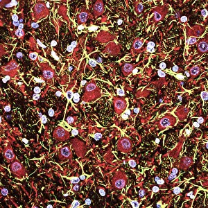
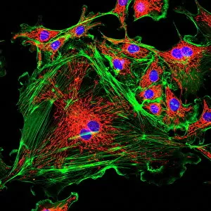
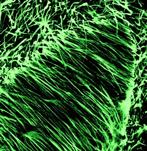
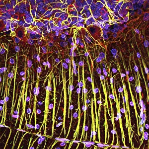
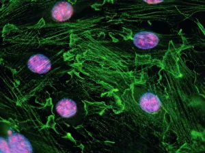
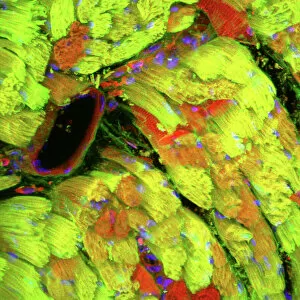
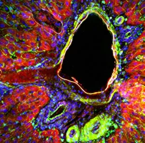
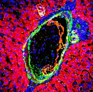
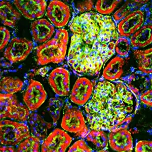
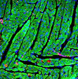
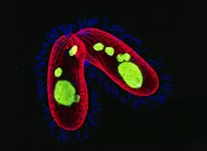
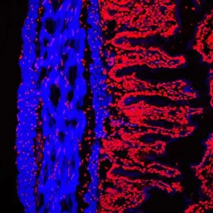
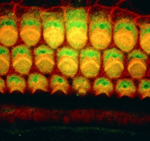
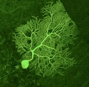
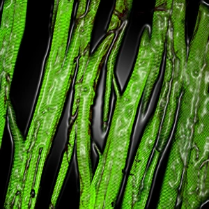
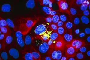
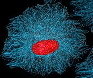
"Exploring the Intricacies of Cerebellum Tissue: A Fascinating Confocal Light Micrograph" This captivating confocal light micrograph unveils the intricate beauty of cerebellum tissue, offering a glimpse into its mesmerizing cell structure. The image showcases glial cells, which play a crucial role in supporting and nourishing nerve cells within the cerebral cortex. The vibrant colors and detailed textures captured by this confocal light micrograph highlight the complexity of these vital brain components. Each glial cell appears as a delicate network, intricately intertwined with neighboring nerve cells to facilitate efficient communication. In another striking view, an immunofluorescent LM reveals fibroblast cell nuclei during cellular division. This dynamic process is reminiscent of uterine cells during childbirth - a testament to the remarkable similarities found across different biological systems. Moving beyond neural tissues, this collection also includes an awe-inspiring confocal light micrograph showcasing heart muscle. The rhythmic pattern and interwoven fibers illustrate the incredible strength and coordination required for proper cardiac function. Additionally, we delve into liver portal triads through two distinct light micrographs. These images shed light on their unique cellular architecture and serve as a reminder of our body's remarkable ability to maintain homeostasis amidst constant metabolic demands. Through these captivating glimpses into various tissues at microscopic levels, we gain deeper insights into their structures and functions. Confocal light microscopy continues to be an invaluable tool in unraveling nature's intricate tapestry at the cellular level.