Fraud Blocker Skip to main content

Cultured Collection

"Cultured: Exploring the Beauty and Complexity of Life's Artistry" Step into a world where art meets science, where beauty intertwines with knowledge

Background imageCultured Collection: Bust of goddess Tanit. Carthaginian art. Sculpture

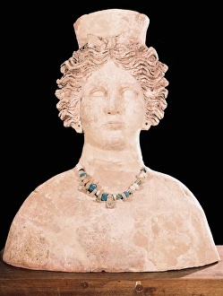
Bust of goddess Tanit. Carthaginian art. Sculpture. SPAIN. BALEARES. IBIZA. Ibiza. Museum of Archaeology of Ibiza and Formentera

Background imageCultured Collection: HeLa cells, light micrograph C017 / 8299

HeLa cells, light micrograph C017 / 8299
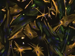
HeLa cells, multiphoton fluorescence micrograph (MFM). The cell nuclei, which contain the cells genetic information, are purple. Microtubules are blue and actin microfilaments are red

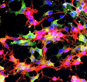
Background imageCultured Collection: Glial stem cell culture, light micrograph

Glial stem cell culture, light micrograph
Glial stem cell culture. Fluorescent light micrograph of glial stem cells producing the protein NG2 (red) as they mature

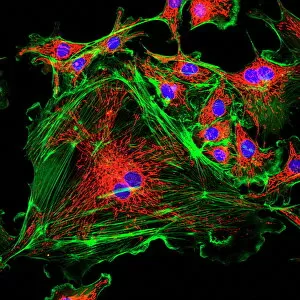
Background imageCultured Collection: Cell structure

Cell structure. Confocal light micrograph of cultured endothelial cells. A fluorescent dye has been used to show the cell structure

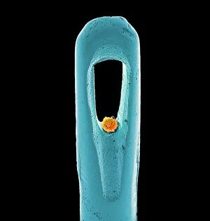
Background imageCultured Collection: Embryonic stem cell and needle, SEM

Embryonic stem cell and needle, SEM
Embryonic stem cell and needle. Coloured scanning electron micrograph (SEM) of an embryonic stem cell (ESC) sitting in the eye of a needle

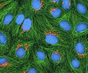
Background imageCultured Collection: HeLa cells, light micrograph C017 / 8298

HeLa cells, light micrograph C017 / 8298
HeLa cells, multiphoton fluorescence micrograph (MFM). The cell nuclei, which contain the cells genetic information, are blue. Golgi bodies, which modify and package proteins, are orange

Background imageCultured Collection: Neural stem cell culture

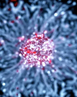
Neural stem cell culture. Fluorescent light micrograph of a group of neural stem cells (neurosphere) in culture

Background imageCultured Collection: HeLa cells, light micrograph C013 / 4774

HeLa cells, light micrograph C013 / 4774
HeLa cells. Multi-photon fluorescence light micrograph of a group of cultured HeLa cells, showing the cell nuclei, which contain the cells genetic information (DNA, red)

Background imageCultured Collection: Nerve cell, SEM

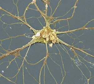
Nerve cell, SEM
Nerve cell. Coloured scanning electron micrograph (SEM) of a neuron (nerve cell). The cell body is the central structure with neurites (long and thin structures) radiating outwards from it

Background imageCultured Collection: Vesicular stomatitis virus, TEM

Vesicular stomatitis virus, TEM
Vesicular stomatitis virus. Coloured transmission electron micrograph (TEM) of vesicular stomatitis virus (VSV) particles (blue) budding from a host cell (red)

Background imageCultured Collection: Vionat of Bar (c.1365-1431). Queen consort of Aragon

Vionat of Bar (c.1365-1431). Queen consort of Aragon
Violant of Bar (c.1365-1431). Queen consort of Aragon by marriage to John I of Aragon, as the second wife of the king. Engraving by Capuz, Historia General de Espana by Father Mariana. Madrid, 1852

Background imageCultured Collection: SPAIN. Kortezubi. Horse (13000 BF). Upper Paleolithic

SPAIN. Kortezubi. Horse (13000 BF). Upper Paleolithic. Magdalenian. Cave

Background imageCultured Collection: Bust of goddess Tanit (terracotta)

Bust of goddess Tanit (terracotta)
CUL4935069 Bust of goddess Tanit (terracotta); (add.info.: Carthaginian art. Sculpture Ibiza. Museum of Archaeology of Ibiza and Formentera); AIC

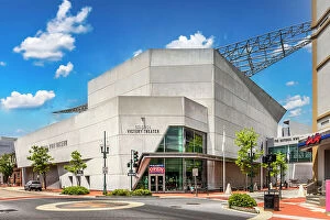
Background imageCultured Collection: Louisiana, New Orleans, The National WWII Museum

Louisiana, New Orleans, The National WWII Museum

Background imageCultured Collection: A Tea Party, c. 1720 (oil on canvas)

A Tea Party, c. 1720 (oil on canvas)
MAN277400 A Tea Party, c.1720 (oil on canvas) by Aken, Joseph van (c.1700-49); 37.4x45.7 cm; Manchester Art Gallery, UK; © Manchester Art Gallery

Background imageCultured Collection: Hush! (The Concert), c. 1875 (oil on canvas)

Hush! (The Concert), c. 1875 (oil on canvas)
2653752 Hush! (The Concert), c.1875 (oil on canvas) by Tissot, James Jacques Joseph (1836-1902); 73.6x112.2 cm; Manchester Art Gallery, UK; © Manchester Art Gallery

Background imageCultured Collection: Phalic dance of type stylized-naturalistic, Mesolithic art (mural painting)

Phalic dance of type stylized-naturalistic, Mesolithic art (mural painting)
CUL4855678 Phalic dance of type stylized-naturalistic, Mesolithic art (mural painting) by Prehistoric; Archaeological Museum of Catalonia, Barcelona, Spain; (add.info.: ILEIDA, El Cogul)

Background imageCultured Collection: DDE-90024833

DDE-90024833
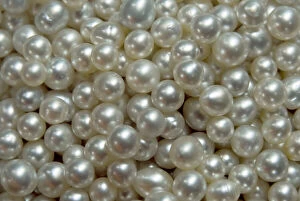
Bowl of pearls cultured from silver-lipped pearl oysters, Raja Ampat Islands, Irian Jaya, West Papua, Indonesia. Date: 12/08/2006

Background imageCultured Collection: Portrait of Marguerite d Angouleme (1492-1549) Queen of Navarre with a dog on her knees

Portrait of Marguerite d Angouleme (1492-1549) Queen of Navarre with a dog on her knees
CHT211469 Portrait of Marguerite d Angouleme (1492-1549) Queen of Navarre with a dog on her knees, facsimile of a sixteenth century drawing, 1848 (colour litho) by French School

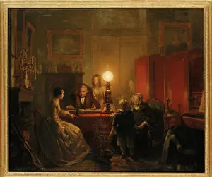
Background imageCultured Collection: A family gathered around a lamplit table, 1854 (oil on canvas)

A family gathered around a lamplit table, 1854 (oil on canvas)
1199807 A family gathered around a lamplit table, 1854 (oil on canvas) by Kiers, Petrus (1807-75); 61.5x73 cm; Private Collection; Photo eChristies Images; Dutch, out of copyright

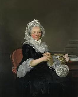
Background imageCultured Collection: Mrs Pearce, 1786 (oil on canvas)

Mrs Pearce, 1786 (oil on canvas)
WAG13339 Mrs Pearce, 1786 (oil on canvas) by Wheatley, Francis (1747-1801); 134.6x10⁹

Background imageCultured Collection: Shakespeares House, Stratford-on-Avon (photo)

Shakespeares House, Stratford-on-Avon (photo)
727516 Shakespeares House, Stratford-on-Avon (photo) by English Photographer, (20th century); Private Collection; (add.info.: Shakespeares House, Stratford-on-Avon)

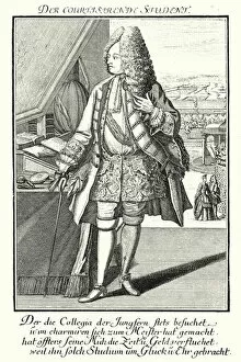
Background imageCultured Collection: The Refined Student (engraving)

The Refined Student (engraving)
3648039 The Refined Student (engraving) by German School, (19th century); Private Collection; (add.info.: The Refined Student)

Background imageCultured Collection: Portrait of Elizabeth, Lady Webster, later Lady Holland

Portrait of Elizabeth, Lady Webster, later Lady Holland
CH441582 Portrait of Elizabeth, Lady Webster, later Lady Holland, in a white dress with her spaniel Pierrot, c.1795 (oil on canvas) by Gauffier

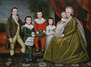
Background imageCultured Collection: Mrs. Noah Smith and Her Children, 1798 (oil on canvas)

Mrs. Noah Smith and Her Children, 1798 (oil on canvas)
5667215 Mrs. Noah Smith and Her Children, 1798 (oil on canvas) by Earl, Ralph (1751-1801); 162.6 x 217.8 cm; Metropolitan Museum of Art, New York

Background imageCultured Collection: Count Francesco Algarotti, 1745 (pastel on vellum)

Count Francesco Algarotti, 1745 (pastel on vellum)
4071451 Count Francesco Algarotti, 1745 (pastel on vellum) by Liotard, Jean-Etienne (1702-89); 42x32.5 cm; Rijksmuseum, Amsterdam

Background imageCultured Collection: At the Sculpture Exhibition, 1895 (oil on canvas)

At the Sculpture Exhibition, 1895 (oil on canvas)
3247951 At the Sculpture Exhibition, 1895 (oil on canvas) by Curran, Charles Courtney (1861-1942); 45.7x55.9 cm; Yale University Art Gallery, New Haven, CT

Background imageCultured Collection: Portrait of a Gentlema, traditionally identified as Sir Robert Sidney (1563-1626

Portrait of a Gentlema, traditionally identified as Sir Robert Sidney (1563-1626
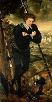
CH249881 Portrait of a Gentlema, traditionally identified as Sir Robert Sidney (1563-1626) later 1st Earl of Leicester (oil on panel) by English School

Background imageCultured Collection: Perspective View of the Salon of the Royal Academy of Painting

Perspective View of the Salon of the Royal Academy of Painting
STC169279 Perspective View of the Salon of the Royal Academy of Painting and Sculpture at the Louvre, Paris (engraving) by French School

Background imageCultured Collection: Society for the Encouragement of Arts, from Ackermanns Microcosm of London

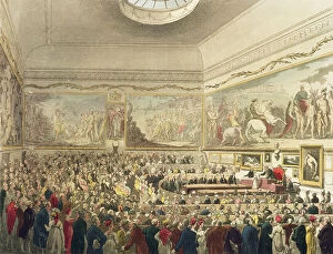
Society for the Encouragement of Arts, from Ackermanns Microcosm of London
STC170342 Society for the Encouragement of Arts, from Ackermanns Microcosm of London, engraved by John Bluck (fl.1791-1831), 1809 (aquatint) by Rowlandson, T. (1756-1827) & Pugin, A.C

Background imageCultured Collection: The Field Family in a Garden, 1869. Creator: Daniel Huntington

The Field Family in a Garden, 1869. Creator: Daniel Huntington
The Field Family in a Garden, 1869

Background imageCultured Collection: Portrait of Madame de Pompadour, ca. 1745. Creator: Charles Nicolas Cochin

Portrait of Madame de Pompadour, ca. 1745. Creator: Charles Nicolas Cochin
Portrait of Madame de Pompadour, ca. 1745

Background imageCultured Collection: Antibiotic action, historical image

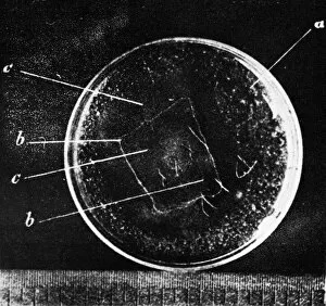
Antibiotic action, historical image
Antibiotic action

Background imageCultured Collection: The Child of Good Breeding from The Complete Works of Beranger, 1836

The Child of Good Breeding from The Complete Works of Beranger, 1836

Background imageCultured Collection: GREECE. Daphni. Dapni Monastery. Nativity (early

GREECE. Daphni. Dapni Monastery. Nativity (early Comnenian period, ca. 1100). Byzantine art. Mosaic

Background imageCultured Collection: Penicillium colony C014 / 4861

Penicillium colony C014 / 4861
Penicillium colony. Colony of Penicillium chrysogenum fungus growing on agar in a petri dish. This fungus produces the antibiotic penicillin G, seen as small droplets on the surface

Background imageCultured Collection: Cultured Old Ladies

Cultured Old Ladies
Two old dears admire an oil painting at an art gallery. Date: 1950s

Background imageCultured Collection: Anthrax antibiotics research

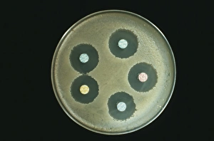
Anthrax antibiotics research. Antibiotic drugs (discs) being tested on anthrax bacteria (Bacillus anthracis) colonies being cultured on an agar growth medium in a petri dish

Background imageCultured Collection: Nerve cell growth

Nerve cell growth. Light micrograph of nerve cells (neurons) with immunofluorescent staining. These cells have been grown in culture

Background imageCultured Collection: Bacteria research

Bacteria research
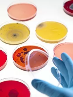
MODEL RELEASED. Bacteria research. Researcher holding an empty petri dish in front of other dishes containing cultured bacteria (dots)

Background imageCultured Collection: Miss Frieda Hempel and M Solito De Solis by Einar Nerman

Miss Frieda Hempel and M Solito De Solis by Einar Nerman
Einar Nermans (1888-1983). Illustration of Miss Frieda Hempel and M Solito De Solis, on stage at the Albert Hall

Background imageCultured Collection: The Museum of Antiquity, in Mustafa Sup鲩eur

The Museum of Antiquity, in Mustafa Sup鲩eur
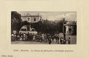
People of Sidi M Hamed, gather in front of the Museum of Antiquity in the area of Mustafa Sup鲩 eur. Date: circa 1910s

Background imageCultured Collection: Nomadic women mince the grain, Ghardaia

Nomadic women mince the grain, Ghardaia
Nomadic women sit in their campsite, with their children, grinding up the grain. Ghardaia, Algeria Date: circa 1910s

Background imageCultured Collection: Famous Diva and accompanist noticed - Oxford Circus Station

Famous Diva and accompanist noticed - Oxford Circus Station
A Famous highbrow Diva (and snooty accompanist) standing on Oxford Circus Underground Station, London - spotted by a couple (and their wailing infant) on the station

Background imageCultured Collection: Tippoo Tib (1837-1905)

Tippoo Tib (1837-1905)
Photograph of Tippoo Tib, then Chief of the Manyema, c.1887. His real name was Hamid ibn Mohammed and he was an African-Arab adventurer and warrior, who controlled a large section of Central Africa

Background imageCultured Collection: Scanning electron micrograph of just-divided HeLa cells

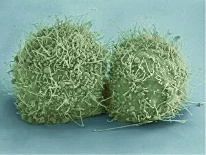
Scanning electron micrograph of just-divided HeLa cells. Zeiss Merlin HR-SEM

Background imageCultured Collection: Multiphoton fluorescence image of HeLa cells

Multiphoton fluorescence image of HeLa cells with cytoskeletal microtubules (magenta) and DNA (cyan)

Background imageCultured Collection: An automatic spinning device to mix tissue culture medium

An automatic spinning device to mix tissue culture medium. Tissue culture medium provides nourishment to growing cells

Background imageCultured Collection: Primary cultures of superior cervical ganglia cells

Primary cultures of superior cervical ganglia cells

Background imageCultured Collection: Scanning electron micrograph of an apoptotic HeLa cell

Scanning electron micrograph of an apoptotic HeLa cell. Zeiss Merlin HR-SEM

Background imageCultured Collection: Directed differentiation of multipotential human neural progenitor cells

Directed differentiation of multipotential human neural progenitor cells
Human neural progenitor cells were isolated under selective culture conditions from the developing human brain and directed through lineage differentiation to GFAP + (glial fibrillary acid protein)

Background imageCultured Collection: Frederick, Prince of Wales and his Sisters, 1733 (oil on canvas)

Frederick, Prince of Wales and his Sisters, 1733 (oil on canvas)
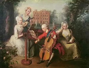
XCF285175 Frederick, Prince of Wales and his Sisters, 1733 (oil on canvas) by Mercier, Philippe (1689-1760); 48x57.78 cm; National Portrait Gallery, London, UK; (add.info.: Frederick Lewis)

Background imageCultured Collection: Viewing at the Royal Academy, c. 1815 (pen, ink and w / c on paper)

Viewing at the Royal Academy, c. 1815 (pen, ink and w / c on paper)
XYC151874 Viewing at the Royal Academy, c.1815 (pen, ink and w/c on paper) by Rowlandson, Thomas (1756-1827); 14.8x24 cm; Yale Center for British Art, Paul Mellon Collection, USA; English

Background imageCultured Collection: George, 3rd Earl Cowper, with the Family of Charles Gore, c. 1775 (oil on canvas)

George, 3rd Earl Cowper, with the Family of Charles Gore, c. 1775 (oil on canvas)
XYC111962 George, 3rd Earl Cowper, with the Family of Charles Gore, c.1775 (oil on canvas) by Zoffany, Johann (1733-1810); 78.7x97.8 cm; Yale Center for British Art, Paul Mellon Collection

Background imageCultured Collection: Italian garden lawn Butchart Gardens Victoria British Columbia Canada

Italian garden lawn Butchart Gardens Victoria British Columbia Canada

Background imageCultured Collection: Neural progenitor cell differentiation C018 / 8758

Neural progenitor cell differentiation C018 / 8758
Neural progenitor cell differentiation. Fluorescence light micrograph of neural progenitor cells that have been grown in a medium that is selective for astrocytes for three weeks

Background imageCultured Collection: Stem cell-derived retinal cells

Stem cell-derived retinal cells. Fluorescence light micrograph of retinal pigment epithelium (RPE) cells that have been derived from human embryonic stem cells (HESC)

Background imageCultured Collection: Stem cell-derived neurons, micrograph

Stem cell-derived neurons, micrograph
Stem cell-derived neurons. Light micrograph of human nerve cells (neurons) that have been derived from induced pluripotent stem cells (IPS). Tuj1 proteins are cyan, and cell nuclei are red

Background imageCultured Collection: HeLa cell, SEM C014 / 0371

HeLa cell, SEM C014 / 0371
HeLa cell. Coloured scanning electron micrograph (SEM) of a HeLa cell (centre) grown in a 3D matrix (background)

Background imageCultured Collection: Biological research F006 / 9086

Biological research F006 / 9086
MODEL RELEASED. Biological research

Background imageCultured Collection: Stem cell-derived astrocyte brain cells

Stem cell-derived astrocyte brain cells
Stem cell-derived nerve cells. Fluorescence light micrograph of astrocyte brain cells that have been derived from neural (nerve) stem cells from a mouse

Background imageCultured Collection: Science mentoring, conceptual artwork

Science mentoring, conceptual artwork
Science mentoring. Conceptual artwork of a senior scientist (right) mentoring a younger one, represented as being cultured in a petri dish (left)

Background imageCultured Collection: Bird flu research C015 / 8803

Bird flu research C015 / 8803
Bird flu research. Researcher harvesting influenza type a H7N9 viruses from cultures grown in eggs. This virus first emerged in the human population in China, in March 2013

Background imageCultured Collection: Bird flu diagnostic kit C015 / 8805

Bird flu diagnostic kit C015 / 8805
Bird flu diagnostic kit. Reagents for diagnosing influenza type a H7N9 infection in humans. This virus first emerged in the human population in China, in March 2013

Background imageCultured Collection: Bird flu research C015 / 8804

Bird flu research C015 / 8804
Bird flu research. Researcher measuring harvested influenza type a H7N9 viruses grown in the lab. This virus first emerged in the human population in China, in March 2013

Background imageCultured Collection: HeLa cells, SEM C014 / 0366

HeLa cells, SEM C014 / 0366
HeLa cells. Coloured scanning electron micrograph (SEM) of HeLa cells (round) grown in a 3D matrix (background)

Background imageCultured Collection: HeLa cells, SEM C014 / 0369

HeLa cells, SEM C014 / 0369
HeLa cells. Coloured scanning electron micrograph (SEM) of HeLa cells (round) grown in a 3D matrix (background)

Background imageCultured Collection: Penicillium colony C014 / 4857

Penicillium colony C014 / 4857
Penicillium colony. Colony of Penicillium chrysogenum fungus growing on agar in a petri dish. This fungus produces the antibiotic penicillin G, seen as small droplets on the surface

Background imageCultured Collection: Hand bacteria C014 / 4839

Hand bacteria C014 / 4839
Hand bacteria. Colony of bacteria that have grown where a hand was pressed onto an agar plate

Background imageCultured Collection: Induced stem cells, light micrograph

Induced stem cells, light micrograph
Induced stem cells. Light micrograph of a cultured colony of induced human pluripotent stem (IPS) cells. Oct-4 (octamer-binding transcription factor 4) is cyan

Background imageCultured Collection: Induced nerve stem cells, micrograph

Induced nerve stem cells, micrograph
Induced nerve stem cells. Fluorescence light micrograph of neural (nerve) stem cells that have been created (induced) from human adult skin fibroblast cells by gene manipulation

Background imageCultured Collection: Stem cell-derived nerve cells

Stem cell-derived nerve cells. Fluorescence light micrograph of neural (nerve) stem cells that have been derived from human embryonic stem cells (HESC)

Background imageCultured Collection: MDCK culture cells, SEM C016 / 8028

MDCK culture cells, SEM C016 / 8028
MDCK cells. Coloured scanning electron micrograph (SEM) of cultured MDCK (Madin-Darby canine kidney) epithelial cells on filter paper (fibres)

Background imageCultured Collection: Microalgae research

Microalgae research. Biologist holding a Petri dish containing cultured diatom microalgae

Background imageCultured Collection: Felix Rey, French virologist

Felix Rey, French virologist. Rey is director of research on the Chikungunya virus research project at the Pasteur Institute, Paris France

Background imageCultured Collection: Chikungunya virus research

Chikungunya virus research. Researcher working with cells cultured from Drosophila sp. fruit flies that have been genetically engineered to produce proteins of the Chikungunya virus

Background imageCultured Collection: Cancer cell, SEM C014 / 0628

Cancer cell, SEM C014 / 0628
Cancer cell. Coloured scanning electron micrograph (SEM) of a carcinoma cancer cell (pink) migrating (metastasising) along a layer of normal epithelial cells (purple)

Background imageCultured Collection: Cancer cell, SEM C014 / 0627

Cancer cell, SEM C014 / 0627
Cancer cell. Coloured scanning electron micrograph (SEM) of a carcinoma cancer cell (yellow) migrating (metastasising) along a layer of normal epithelial cells (brown)

Background imageCultured Collection: HeLa cells, SEM C017 / 8305

HeLa cells, SEM C017 / 8305
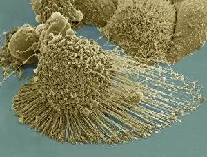
HeLa cells, coloured scanning electron micrograph (SEM). The cells have just replicated. HeLa cells were the first continuously cultured cell line

Background imageCultured Collection: HeLa cell dying, SEM C017 / 8304

HeLa cell dying, SEM C017 / 8304
HeLa cell dying, coloured scanning electron micrograph (SEM). The cell in the foreground is undergoing apoptosis, or programmed cell death. Apoptosis occurs when a cell becomes old or damaged

Background imageCultured Collection: Microbiology laboratory C013 / 7245

Microbiology laboratory C013 / 7245
Microbiology laboratory. Collection of glass bottles containing cultured bacterial samples in a microbiology lab

Background imageCultured Collection: Microbiology laboratory

Microbiology laboratory. Collection of glass bottles containing cultured bacterial samples in a microbiology lab

Background imageCultured Collection: Keratinocyte skin cells, SEM C013 / 4781

Keratinocyte skin cells, SEM C013 / 4781
Keratinocyte skin cells. Coloured scanning electron micrograph (SEM) of human keratinocyte skin cells. These epithelial cells produce keratin in the skins outermost layers

Background imageCultured Collection: Keratinocyte skin cells, SEM C013 / 4780

Keratinocyte skin cells, SEM C013 / 4780
Keratinocyte skin cells. Coloured scanning electron micrograph (SEM) of human keratinocyte skin cells. These epithelial cells produce keratin in the skins outermost layers


All products are expertly crafted, using premium materials, tailored to your specifications and promptly shipped

"Cultured: Exploring the Beauty and Complexity of Life's Artistry" Step into a world where art meets science, where beauty intertwines with knowledge. From ancient civilizations to modern breakthroughs, the concept of "cultured" takes on new meaning as we delve into the wonders of human creativity and scientific exploration. Intricate sculptures like the Bust of goddess Tanit transport us back to Carthaginian times, showcasing their mastery in capturing divine beauty through art. The attention to detail and craftsmanship displayed in these pieces is truly awe-inspiring. But our journey doesn't stop there; it delves deeper into the microscopic realm. Witnessing HeLa cells under a light micrograph reveals their intricate structures, reminding us that even at this minuscule level, life holds immense complexity and wonder. Glial stem cell cultures further highlight how different types of cells can be nurtured and grown in controlled environments for research purposes. Embryonic stem cells take center stage alongside a needle in an SEM image, symbolizing both hope for medical advancements and ethical debates surrounding their use. These versatile cells hold tremendous potential for regenerative medicine but also raise questions about ethics and boundaries. Neural stem cell cultures demonstrate our ability to cultivate specific types of brain cells outside their natural environment – a testament to humanity's relentless pursuit of understanding our own minds. Nerve cells captured through SEM imagery showcase their intricate branching patterns that enable communication within our nervous system. Venturing beyond human biology, we encounter Vesicular stomatitis virus under TEM – an insight into microbial worlds that shape our existence. Bacterial cultures remind us that life thrives not only within ourselves but also all around us – invisible yet impactful. Finally, plant biotechnology emerges as another facet expression. Through genetic engineering techniques, scientists have harnessed nature's potential by modifying plants' genetic makeup for various purposes such as increased crop yield or disease resistance.

© Copyright Media Storehouse, All Rights Reserved 2003 - 2024
Facebook     Pinterest     100% Payment Secure     Reviews IO