Fraud Blocker Skip to main content

Cytology Collection (page 5)

Cytology: Exploring the Intricate World of Cells Budding yeast cell: Witness the remarkable process of cellular reproduction as a budding yeast cell emerges

Background imageCytology Collection: Conceptual image of cytoskeleton

Conceptual image of cytoskeleton. The cytoskeleton is a cellular scaffolding or skeleton contained within a cells cytoplasm, and is present in all cells

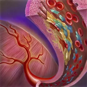
Background imageCytology Collection: Artery showing atherosclerotic plaque, platelets and red blood cells

Artery showing atherosclerotic plaque, platelets and red blood cells
Close-up of artery showing atherosclerotic plaque, platelets and red blood cells

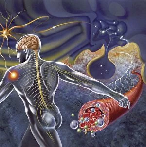
Background imageCytology Collection: Schematic of the hypothalamus receiving nerve impulses from the body

Schematic of the hypothalamus receiving nerve impulses from the body and sending messages to the circulatory and nervous system

Background imageCytology Collection: Artistic representation of the immune systems reaction to bacteria invading the tissues

Artistic representation of the immune systems reaction to bacteria invading the tissues

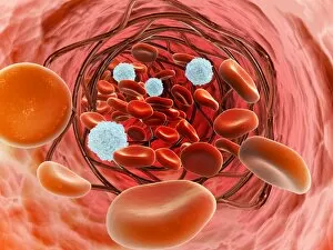
Background imageCytology Collection: Arteriole with red blood cells, white blood cells and oxygen

Arteriole with red blood cells, white blood cells and oxygen

Background imageCytology Collection: Conceptual image of synaptic transmission

Conceptual image of synaptic transmission

Background imageCytology Collection: Conceptual image of helicobacter

Conceptual image of helicobacter. Helicobacter is a genus of gram-negative bacteria possessing a characteristic helix shape

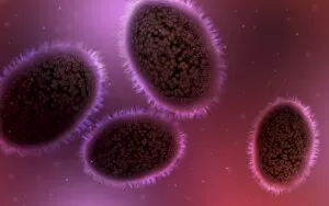
Background imageCytology Collection: Microscopic view of samllpox

Microscopic view of samllpox

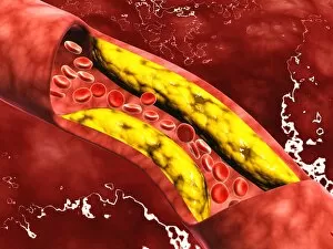
Background imageCytology Collection: Microscopic view of fat plaque inside the artery

Microscopic view of fat plaque inside the artery

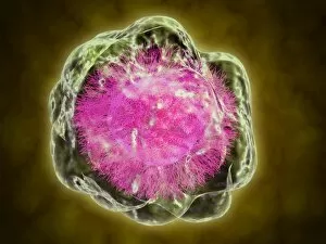
Background imageCytology Collection: Microscopic view of human cell

Microscopic view of human cell

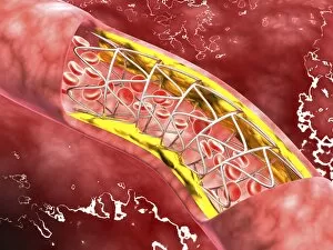
Background imageCytology Collection: Microscopic view of blood flow inside an artery with stent deployment

Microscopic view of blood flow inside an artery with stent deployment

Background imageCytology Collection: Microscopic view of a bipolar neuron

Microscopic view of a bipolar neuron. A bipolar cell is a type of neuron which has two extensions. Bipolar cells are specialized sensory neurons for the transmission of special senses

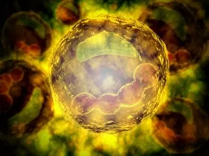
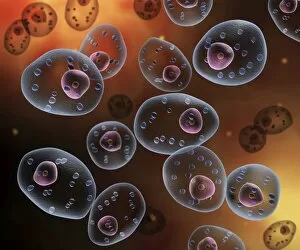
Background imageCytology Collection: The interior of an eukaryotic cell

The interior of an eukaryotic cell. This is the most common generic type of cell and its present in all mammals. In the center is the nucleus (the perforated sphere)

Background imageCytology Collection: Insertion of stent into atherosclerotic artery

Insertion of stent into atherosclerotic artery

Background imageCytology Collection: Conceptual image of mast cells

Conceptual image of mast cells

Background imageCytology Collection: Conceptual image of a tumor in human brain

Conceptual image of a tumor in human brain

Background imageCytology Collection: Artery cross-section with blood flow, fat plaque and stent deployment

Artery cross-section with blood flow, fat plaque and stent deployment
Microscopic view of an artery cross-section with blood flow, fat plaque and stent deployment

Background imageCytology Collection: Conceptual image of Encephalitis

Conceptual image of Encephalitis. Encephalitis can cause flu-like symptoms, such as a fever or severe headaches. It can also cause confused thinking, seizures, or problems with senses
Background imageCytology Collection: Conceptual image of red blood cells with platelets

Conceptual image of red blood cells with platelets

Background imageCytology Collection: Microscopic view of HIV virus inside the lungs

Microscopic view of HIV virus inside the lungs

Background imageCytology Collection: Microscopic view of plasma cell inside blood vessel

Microscopic view of plasma cell inside blood vessel

Background imageCytology Collection: Close-up view of the synapse in the nervous system

Close-up view of the synapse in the nervous system. A synapse is a structure that permits a neuron (or nerve cell) to pass an electrical or chemical signal to another cell

Background imageCytology Collection: Inside view of the artery with red blood cells

Inside view of the artery with red blood cells

Background imageCytology Collection: Microscopic view of a mast cell

Microscopic view of a mast cell. Mast cells are found resident in tissues throughout the body, particularly in association with structures such as blood vessels and nerves

Background imageCytology Collection: Microscopic view of birch tree pollen

Microscopic view of birch tree pollen. Birch tree pollen is considered to be an important allergenic tree pollen

Background imageCytology Collection: Microscopic view of stem cell development

Microscopic view of stem cell development. In adult organisms, stem cells and progenitor cells act as a repair system for the body, replenishing adult tissues

Background imageCytology Collection: Conceptual image of mitosis

Conceptual image of mitosis. Mitosis is the process in the cell cycle by which a cell duplicates into two genetically identical sets of chromosomes

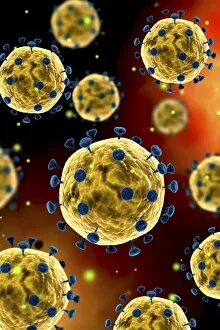
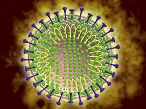
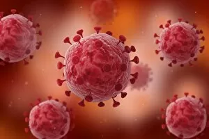
Background imageCytology Collection: Microscopic view of coronavirus

Microscopic view of coronavirus

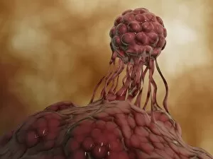
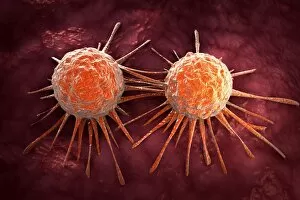
Background imageCytology Collection: Conceptual image of cancer virus

Conceptual image of cancer virus

Background imageCytology Collection: Conceptual image of red blood cells with fibrin

Conceptual image of red blood cells with fibrin

Background imageCytology Collection: Microscopic view of a group of macrophages

Microscopic view of a group of macrophages. Macrophages contribute to tumor growth and progression. Attracted to oxygen-starved (hypoxic) and necrotic tumor cells they promote chronic inflammation

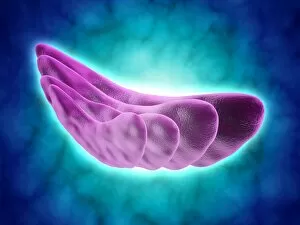
Background imageCytology Collection: Conceptual image of the Golgi apparatus

Conceptual image of the Golgi apparatus. The Golgi apparatus is an organelle found in most eukaryotic cells

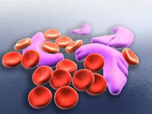
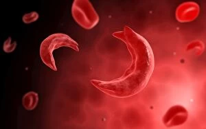
Background imageCytology Collection: Conceptual image of sickle cell anemia

Conceptual image of sickle cell anemia. Sickle cell anemia is a disease in which your body produces abnormally shaped red blood cells. The cells are shaped like a crescent or sickle

Background imageCytology Collection: Nerve with myelin sheath, seen in lower right, connects with muscle

Nerve with myelin sheath, seen in lower right, connects with muscle. Blood vessel and immune cells are seen in the center and upper right of image

Background imageCytology Collection: Conceptual image of the coronavirus

Conceptual image of the coronavirus. Coronaviruses primarily infect the upper respiratory and gastrointestinal tract

Background imageCytology Collection: Microscopic view of virus

Microscopic view of virus

Background imageCytology Collection: Conceptual image of endoplasmic reticulum around a cell nucleus

Conceptual image of endoplasmic reticulum around a cell nucleus. Endoplasmic reticulum is an organelle that forms a continuous membrane system of flattened sacs within the cytoplasm of a eukaryotic

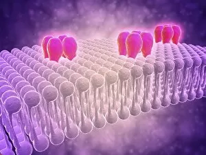
Background imageCytology Collection: Conceptual image of GABA receptors

Conceptual image of GABA receptors. The GABA receptors are a class of receptors that respond to the neurotransmitter gamma-aminobutyric acid

Background imageCytology Collection: Microscopic view of herpes virus

Microscopic view of herpes virus

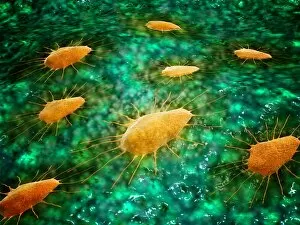
Background imageCytology Collection: Conceptual image of Trypanosoma

Conceptual image of Trypanosoma

Background imageCytology Collection: Conceptual image of influenza causing flu virus

Conceptual image of influenza causing flu virus

Background imageCytology Collection: 3D rendering of macrophage phagocytosis

3D rendering of macrophage phagocytosis

Background imageCytology Collection: Microscopic view of phagocytic macrophages

Microscopic view of phagocytic macrophages, which are involved in the immune response within the body

Background imageCytology Collection: Group of Escherichia coli bacteria cells, known as E. Coli

Group of Escherichia coli bacteria cells, known as E. Coli
Group of Escherichia coli bacteria cells, commonly known as E. Coli. E. coli is a common type of bacteria that can get into food, like beef and vegetables

Background imageCytology Collection: Microscopic view of human anitbodies with red blood cells

Microscopic view of human anitbodies with red blood cells. Human antibodies are the Y-shaped proteins used by the immune system to defend against foreign objects like bacteria and viruses

Background imageCytology Collection: Conceptual image of a neuron ligntning signal passing

Conceptual image of a neuron ligntning signal passing

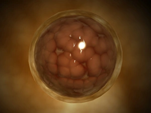
Background imageCytology Collection: Microscopic view of a blastula during pregnancy

Microscopic view of a blastula during pregnancy. After the cleavage has produced over 100 cells, the embryo is called a blastula

Background imageCytology Collection: Microscopic view of sicke cells causing anemia disease

Microscopic view of sicke cells causing anemia disease


All Professionally Made to Order for Quick Shipping

Cytology: Exploring the Intricate World of Cells Budding yeast cell: Witness the remarkable process of cellular reproduction as a budding yeast cell emerges, paving the way for new life. T lymphocytes and cancer cell, SEM C001 / 1679: Delve into the battle between our immune system's T lymphocytes and cancer cells, captured in stunning detail through scanning electron microscopy. Anaesthetic inhibiting an ion channel C015 / 6718: Uncover the secrets behind anesthesia as we observe its impact on ion channels within cells, revealing how it alters our perception of pain. Glial cells, confocal light micrograph: Immerse yourself in the intricate network of glial cells that support and protect neurons in our nervous system, visualized using confocal light microscopy. HeLa cells, light micrograph C017 / 8299: Explore one of the most famous cell lines - HeLa cells - under a microscope to gain insights into their structure and function within biomedical research. Rough endoplasmic reticulum, TEM: Journey deep inside a cell to witness the rough endoplasmic reticulum at work – a vital organelle involved in protein synthesis – revealed through transmission electron microscopy. Glial stem cell culture, light micrograph: Step into the realm of regenerative medicine as we observe glial stem cells cultured in vitro with hopes of unlocking their potential for repairing damaged neural tissue. Dendritic cells, artwork: Marvel at an artistic representation showcasing dendritic cells' crucial role as sentinels of our immune system – capturing antigens and presenting them to other immune cells for recognition and response. Cell structure: Embark on a visual journey exploring various cellular structures that form the building blocks of life itself – from membranes to cytoplasm to nuclei – highlighting their diverse functions.