Fraud Blocker Skip to main content

Cytology Collection (page 7)

Cytology: Exploring the Intricate World of Cells Budding yeast cell: Witness the remarkable process of cellular reproduction as a budding yeast cell emerges

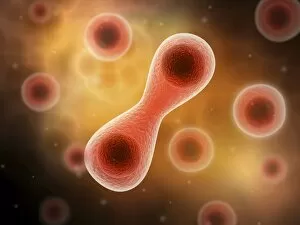
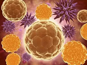
Background imageCytology Collection: Microscopic view of cell division

Microscopic view of cell division

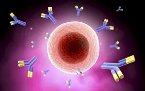
Background imageCytology Collection: Microscopic view of cell and antibody

Microscopic view of cell and antibody

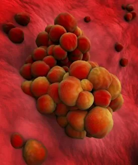
Background imageCytology Collection: Microscopic view of tumor

Microscopic view of tumor

Background imageCytology Collection: Microscopic view of animal cell nucleus

Microscopic view of animal cell nucleus

Background imageCytology Collection: Conceptual image of flagellate bacterium

Conceptual image of flagellate bacterium

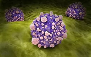
Background imageCytology Collection: Microscopic view of pancreatic cancer cell

Microscopic view of pancreatic cancer cell

Background imageCytology Collection: Microscopic view of male sperm cells

Microscopic view of male sperm cells

Background imageCytology Collection: Microscopic view of a mast cell found in connective tissue

Microscopic view of a mast cell found in connective tissue that contains numerous basophilic granules and releases substances such as heparin and histamine in response to injury

Background imageCytology Collection: Microscopic view of human B-cells

Microscopic view of human B-cells which play a large role in the immune response system

Background imageCytology Collection: Microscopic view showing bone structure of sperm

Microscopic view showing bone structure of sperm

Background imageCytology Collection: Malria spores in the human blood stream

Malria spores in the human blood stream
The malaria spores move from the mosquito to the human blood stream. the spores divide many times and produce large numbers of spores

Background imageCytology Collection: Microscopic view of a multipolar neuron

Microscopic view of a multipolar neuron. Multipolar neurons possess a single axon and many dendrites

Background imageCytology Collection: Conceptual image of a ubiquitous virus

Conceptual image of a ubiquitous virus. A ubiquitous virus is contagious in early childhood through the respiratory tract

Background imageCytology Collection: Microscopic view of Henipavirus

Microscopic view of Henipavirus. Henipavirus is a established group of paramyxoviruses comprising the Hendra virus, Nipah virus and Cedar virus

Background imageCytology Collection: Microscopic view of red blood cells flowing inside lungs

Microscopic view of red blood cells flowing inside lungs

Background imageCytology Collection: Microscipic view of pancreatic cancer cells

Microscipic view of pancreatic cancer cells

Background imageCytology Collection: Illustration of action potential of a nerve cell

Illustration of action potential of a nerve cell

Background imageCytology Collection: Conceptual image of vibrio cholerae causing cholera

Conceptual image of vibrio cholerae causing cholera

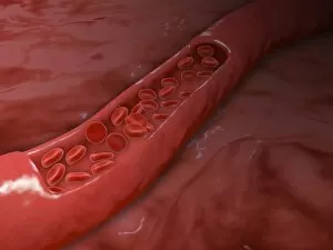
Background imageCytology Collection: Artery cross section with red blood cell flow

Artery cross section with red blood cell flow

Background imageCytology Collection: Conceptual image of lysosome

Conceptual image of lysosome. Lysosomes are cellular organelles that contain acid hydrolase enzymes that break down waste materials and cellular debris

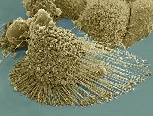
Background imageCytology Collection: Scanning electron micrograph of an apoptotic HeLa cell

Scanning electron micrograph of an apoptotic HeLa cell. Zeiss Merlin HR-SEM

Background imageCytology Collection: Conceptual image of a neuron

Conceptual image of a neuron

Background imageCytology Collection: Microscopic view of a single male sperm cell

Microscopic view of a single male sperm cell

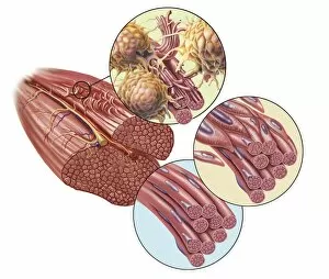
Background imageCytology Collection: Torn muscle fibers with healing stages surrounding

Torn muscle fibers with healing stages surrounding. 1. Macrophage clean-up. 2. New cells migrate to repair. 3. Cells differentiate to become muscle cells

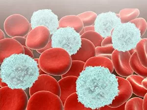
Background imageCytology Collection: Red blood cells with white blood cells

Red blood cells with white blood cells

Background imageCytology Collection: Stochastic gene expression, illustration C018 / 0906

Stochastic gene expression, illustration C018 / 0906
Stochastic gene expression, illustration. Every cell in an organism contains every single gene that makes up the organisms genome. However, they are not all active (expressed) in each cell

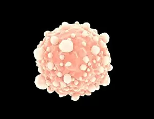
Background imageCytology Collection: Cancer cell, artwork C016 / 9843

Cancer cell, artwork C016 / 9843
Cancer cell. Computer artwork of a cancerous (malignant) cell (top left) in tissue. The uneven surface and cytoplasmic projections are characteristic of cancer cells

Background imageCytology Collection: Biological research F006 / 7189

Biological research F006 / 7189
Biological research

Background imageCytology Collection: Mitochondria, artwork

Mitochondria, artwork
Mitochondria, computer artwork

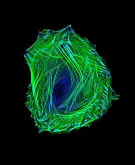
Background imageCytology Collection: Embryonic smooth muscle cell C018 / 8595

Embryonic smooth muscle cell C018 / 8595
Embryonic smooth muscle cell, immunofluorescence micrograph. Actin filaments are green, the protein vinculin is light blue. Actin is part of the cytoskeleton

Background imageCytology Collection: Eosinophil white blood cell, TEM C014 / 1438

Eosinophil white blood cell, TEM C014 / 1438
Eosinophil white blood cell. Transmission electron micrograph (TEM) of a section through an eosinophil. Eosinophils, like all white blood cells, are part of the bodys immune system

Background imageCytology Collection: Bladder lining, TEM C014 / 1473

Bladder lining, TEM C014 / 1473
Bladder lining. Transmission electron micrograph (TEM) of a section through the epithelial cells that line of a human bladder, showing large flat vesicles (large, blue)

Background imageCytology Collection: Intestinal gland cell, TEM C014 / 1441

Intestinal gland cell, TEM C014 / 1441
Intestinal gland cell. Transmission electron micrograph (TEM) of a section through an undifferentiated cell from the intestinal tract, showing extensive rough endoplasmic reticulum (ER)

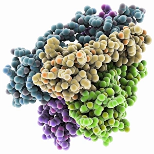
Background imageCytology Collection: Potassium ion channel protein structure

Potassium ion channel protein structure. Molecular model of a KcsA potassium ion (K+) channel from Streptomyces lividans bacteria

Background imageCytology Collection: Potassium ion channel beta subunit

Potassium ion channel beta subunit. Molecular model showing the structure a beta subunit of a voltage-dependent potassium (K+) channel

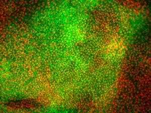
Background imageCytology Collection: Stem cell-derived retinal cells

Stem cell-derived retinal cells. Fluorescence light micrograph of retinal pigment epithelium (RPE) cells that have been derived from human embryonic stem cells (HESC)

Background imageCytology Collection: KCNQ ion channel protein structure

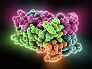
KCNQ ion channel protein structure. Molecular model showing the protein structure of an ion channel domain. Ion channels are membrane-spanning proteins that form a pathway for the movement of

Background imageCytology Collection: Potassium ion channel cavity structure

Potassium ion channel cavity structure. Molecular model showing the structure of a cavity formed by potassium ion channel proteins

Background imageCytology Collection: Stem cell-derived neurons, micrograph

Stem cell-derived neurons, micrograph
Stem cell-derived neurons. Light micrograph of human nerve cells (neurons) that have been derived from induced pluripotent stem cells (IPS). Tuj1 proteins are cyan, and cell nuclei are red

Background imageCytology Collection: ATP synthase molecule C014 / 0880

ATP synthase molecule C014 / 0880
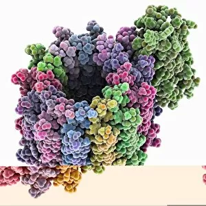
ATP synthase molecule. Molecular model showing the structure of ATP synthase (ATPase) subunit C. ATPase is an important enzyme that provides energy for cells through the synthesis of adenosine

Background imageCytology Collection: Astrocyte brain cells, light micrograph

Astrocyte brain cells, light micrograph
Astrocyte brain cells. Fluorescence light micrograph of primary astrocyte cells from the brain of a mouse. Astrocytes have numerous branches of connective tissue that provide support
Background imageCytology Collection: HeLa cell, SEM C014 / 0371

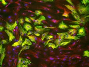
HeLa cell, SEM C014 / 0371
HeLa cell. Coloured scanning electron micrograph (SEM) of a HeLa cell (centre) grown in a 3D matrix (background). HeLa cells are a continuously cultured cell line of immortal human cancer cells

Background imageCytology Collection: Cancer cell, artwork F006 / 2311

Cancer cell, artwork F006 / 2311
Cancer cell, computer artwork

Background imageCytology Collection: Cancer cell, artwork F006 / 2309

Cancer cell, artwork F006 / 2309
Cancer cell, computer artwork

Background imageCytology Collection: Cancer cell, artwork F006 / 2308

Cancer cell, artwork F006 / 2308
Cancer cell, computer artwork

Background imageCytology Collection: Human fat cells, artwork F006 / 2275

Human fat cells, artwork F006 / 2275
Human fat cells, computer artwork

Background imageCytology Collection: Human fat cells, artwork F006 / 2274

Human fat cells, artwork F006 / 2274
Human fat cells, computer artwork

Background imageCytology Collection: Animal cells, artwork F006 / 2225

Animal cells, artwork F006 / 2225
Animal cells, computer artwork


All Professionally Made to Order for Quick Shipping

Cytology: Exploring the Intricate World of Cells Budding yeast cell: Witness the remarkable process of cellular reproduction as a budding yeast cell emerges, paving the way for new life. T lymphocytes and cancer cell, SEM C001 / 1679: Delve into the battle between our immune system's T lymphocytes and cancer cells, captured in stunning detail through scanning electron microscopy. Anaesthetic inhibiting an ion channel C015 / 6718: Uncover the secrets behind anesthesia as we observe its impact on ion channels within cells, revealing how it alters our perception of pain. Glial cells, confocal light micrograph: Immerse yourself in the intricate network of glial cells that support and protect neurons in our nervous system, visualized using confocal light microscopy. HeLa cells, light micrograph C017 / 8299: Explore one of the most famous cell lines - HeLa cells - under a microscope to gain insights into their structure and function within biomedical research. Rough endoplasmic reticulum, TEM: Journey deep inside a cell to witness the rough endoplasmic reticulum at work – a vital organelle involved in protein synthesis – revealed through transmission electron microscopy. Glial stem cell culture, light micrograph: Step into the realm of regenerative medicine as we observe glial stem cells cultured in vitro with hopes of unlocking their potential for repairing damaged neural tissue. Dendritic cells, artwork: Marvel at an artistic representation showcasing dendritic cells' crucial role as sentinels of our immune system – capturing antigens and presenting them to other immune cells for recognition and response. Cell structure: Embark on a visual journey exploring various cellular structures that form the building blocks of life itself – from membranes to cytoplasm to nuclei – highlighting their diverse functions.