Fraud Blocker Skip to main content

Dissection Collection (page 3)

"Unveiling the Wonders Within

Background imageDissection Collection: Experiment on frogs by Louis Galvani (1737-98) late 18th century (engraving) (b / w photo)

Experiment on frogs by Louis Galvani (1737-98) late 18th century (engraving) (b / w photo)
XIR217898 Experiment on frogs by Louis Galvani (1737-98) late 18th century (engraving) (b/w photo) by French School, (18th century); Private Collection; (add.info.: experiment on frogs; ); French

Background imageDissection Collection: Haruspice consulting the entrails of a bull (marble) (see 261376 amd 261378)

Haruspice consulting the entrails of a bull (marble) (see 261376 amd 261378)
ALI261377 Haruspice consulting the entrails of a bull (marble) (see 261376 amd 261378) by Roman, (4th century AD); Louvre, Paris, France; (add.info.: Haruspice, also known as Aruspice, Haruspex)

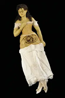
Background imageDissection Collection: Anatomical model of a pregnant woman (wax & textiles)

Anatomical model of a pregnant woman (wax & textiles)
1123883 Anatomical model of a pregnant woman (wax & textiles) by English School, (19th century); Private Collection; (add.info)

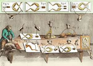
Background imageDissection Collection: Experiment on frogs, illustration from De viribus electricitatis in motu

Experiment on frogs, illustration from De viribus electricitatis in motu
XIR217897 Experiment on frogs, illustration from De viribus electricitatis in motu muscumari (engraving) (b/w photo) by French School, (18th century); Bibliotheque Nationale, Paris

Background imageDissection Collection: Doll showing the anatomy of the human body (ivory)

Doll showing the anatomy of the human body (ivory)
XIR118564 Doll showing the anatomy of the human body (ivory) by German School, (17th century); Museo Nazionale Dell Arte Sanitaria, Rome, Italy; German, out of copyright

Background imageDissection Collection: The Heart, facsimile of the Windsor book (pen and ink on paper)

The Heart, facsimile of the Windsor book (pen and ink on paper)
CHT164556 The Heart, facsimile of the Windsor book (pen and ink on paper) by Vinci, Leonardo da (1452-1519) (after); Bibliotheque des Arts Decoratifs, Paris, France; eArchives Charmet; Italian

Background imageDissection Collection: Fol. 38v The Heart, from Traite de Medecine by Aldebrande de Florence

Fol. 38v The Heart, from Traite de Medecine by Aldebrande de Florence
XIR159270 Fol.38v The Heart, from Traite de Medecine by Aldebrande de Florence, 1356 (vellum) by French School, (14th century); Biblioteca del Palacio Nacional de Ajuda, Lisbon, Portugal; French

Background imageDissection Collection: The Anatomy Lesson (pen & ink)

The Anatomy Lesson (pen & ink)
QED32742 The Anatomy Lesson (pen & ink) by Passarotti, Bartolomeo (1529-92); Louvre, Paris, France; Italian, out of copyright

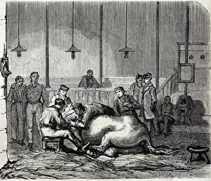
Background imageDissection Collection: The National veterinary school of Alfort - Les grandes ecole de france - ecole

The National veterinary school of Alfort - Les grandes ecole de france - ecole
XEE4161516 The National veterinary school of Alfort - Les grandes ecole de france - ecole veterinaire de maison alfort en 1866 - la salle de dissection - Engraving in " Le monde

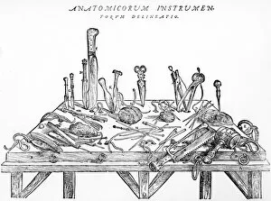
Background imageDissection Collection: Instruments for Dissections, illustration from De Humani Corporis Fabrica

Instruments for Dissections, illustration from De Humani Corporis Fabrica
867832 Instruments for Dissections, illustration from De Humani Corporis Fabrica (engraving) by Vesalius, Andreas (1514-64); Private Collection; eArchives Charmet; Belgian, out of copyright

Background imageDissection Collection: Midwifery, illustration from Encyclopedia Britannica, published in Edinburgh

Midwifery, illustration from Encyclopedia Britannica, published in Edinburgh
DGC398035 Midwifery, illustration from Encyclopedia Britannica, published in Edinburgh, 1798 (copperplate engraving) by Bell, Alexander (fl)

Background imageDissection Collection: Anatomical model of an old mans head (wax)

Anatomical model of an old mans head (wax)
BEN360192 Anatomical model of an old mans head (wax) by Zumbo, Gaetano (1656-1701); Museo della Specola, Florence, Italy; (add.info)

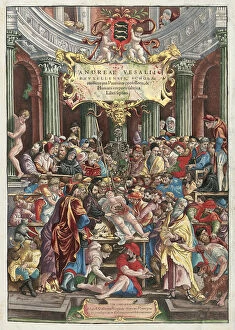
Background imageDissection Collection: Frontispiece to De Humani Corporis Fabrica Libri Septem

Frontispiece to De Humani Corporis Fabrica Libri Septem
CH466697 Frontispiece to De Humani Corporis Fabrica Libri Septem by Andreas Vesalius (1514-64), published by Johannes Oporinus, Basel, June 1543 (colour woodcut) (see also 233019) by Venetian School

Background imageDissection Collection: Experiment on frogs, illustration from De viribus electricitatis in motu

Experiment on frogs, illustration from De viribus electricitatis in motu
XIR232085 Experiment on frogs, illustration from De viribus electricitatis in motu muscumari (engraving) (later colouration) by French School, (18th century); Bibliotheque Nationale, Paris

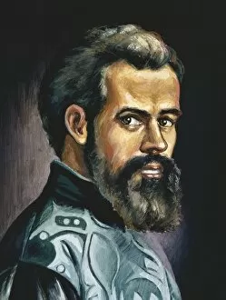
Background imageDissection Collection: Portrait of Andreas Vesalius (1514-64) (oil on canvas)

Portrait of Andreas Vesalius (1514-64) (oil on canvas)
CHT164194 Portrait of Andreas Vesalius (1514-64) (oil on canvas) by Tintoretto, Jacopo Robusti (1518-94) (after); Academie de Medecine, Paris

Background imageDissection Collection: The Heart, plate from Anatomy of the Visceras, dissected

The Heart, plate from Anatomy of the Visceras, dissected
CHT164178 The Heart, plate from Anatomy of the Visceras, dissected, painted and engraved by Gautier (colour engraving) by Gautier D Agoty

Background imageDissection Collection: The Heart and the Circulation, facsimile of the Windsor book (pen and ink on paper)

The Heart and the Circulation, facsimile of the Windsor book (pen and ink on paper)
CHT164554 The Heart and the Circulation, facsimile of the Windsor book (pen and ink on paper) by Vinci, Leonardo da (1452-1519) (after); Bibliotheque des Arts Decoratifs, Paris

Background imageDissection Collection: Laboratory of comparative anatomy (engraving)

Laboratory of comparative anatomy (engraving)
5998509 Laboratory of comparative anatomy (engraving) by Gelhay, Edouard (1856-1939); Private Collection; (add.info.: Laboratory of comparative anatomy)

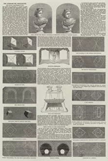
Background imageDissection Collection: The Stereoscope, Pseudoscope, and Solid Daguerreotypes (engraving)

The Stereoscope, Pseudoscope, and Solid Daguerreotypes (engraving)
1607279 The Stereoscope, Pseudoscope, and Solid Daguerreotypes (engraving) by English School, (19th century); Private Collection; (add.info.: The Stereoscope, Pseudoscope, and Solid Daguerreotypes)

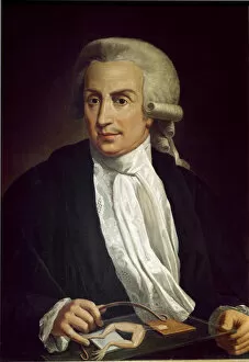
Background imageDissection Collection: Luigi Galvani with frog specimen (oil on canvas)

Luigi Galvani with frog specimen (oil on canvas)
LRI3725846 Luigi Galvani with frog specimen (oil on canvas) by Italian School, (18th century); Universita di Bologna, Italy; (add.info)

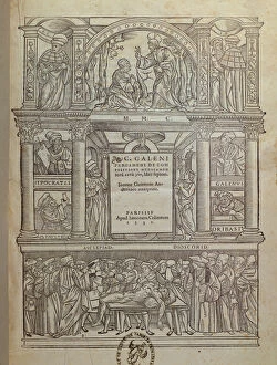
Background imageDissection Collection: Title page of a book by Galen (c. 131-c. 201), published in Paris, 1530 (engraving)

Title page of a book by Galen (c. 131-c. 201), published in Paris, 1530 (engraving)
CHT164175 Title page of a book by Galen (c.131-c.201), published in Paris, 1530 (engraving) by French School, (16th century); Bibliotheque de la Faculte de Medecine, Paris

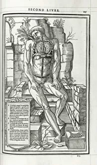
Background imageDissection Collection: Anatomical illustration, 1546 (woodcut)

Anatomical illustration, 1546 (woodcut)
985877 Anatomical illustration, 1546 (woodcut) by French School, (16th century); 37.2x24.6 cm; Private Collection; (add.info.: Anatomical illustration)

Background imageDissection Collection: The Author Conducting an Anatomical Dissection in a Surgical Theatre, 1543 (woodcut)

The Author Conducting an Anatomical Dissection in a Surgical Theatre, 1543 (woodcut)
985793 The Author Conducting an Anatomical Dissection in a Surgical Theatre, 1543 (woodcut) by Italian School, (16th century); 42.1x29 (folio) cm; Private Collection; (add.info)

Background imageDissection Collection: The Gross Clinic, 1876. Creator: Thomas Eakins

The Gross Clinic, 1876. Creator: Thomas Eakins
The Gross Clinic, 1876

Background imageDissection Collection: Male head in profile to left with muscles exposed, 1660. Creator: Wenceslaus Hollar

Male head in profile to left with muscles exposed, 1660. Creator: Wenceslaus Hollar
Male head in profile to left with muscles exposed, 1660

Background imageDissection Collection: The Dissecting Room, ca. 1838. Creator: T. C. Wilson

The Dissecting Room, ca. 1838. Creator: T. C. Wilson
The Dissecting Room, ca. 1838

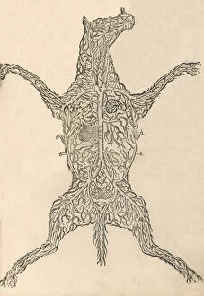
Background imageDissection Collection: Anatomia del Cavallo. Infirmita del Cavallo, 1602. Creator: Carlo Ruini

Anatomia del Cavallo. Infirmita del Cavallo, 1602. Creator: Carlo Ruini
Anatomia del Cavallo. Infirmita del Cavallo, 1602

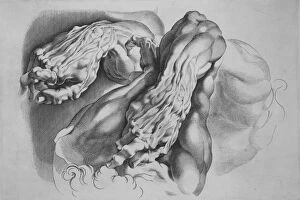
Background imageDissection Collection: Three Studies of Ecorches, after 1640. after 1640. Creator: Paulus Pontius

Three Studies of Ecorches, after 1640. after 1640. Creator: Paulus Pontius
Three Studies of Ecorches, after 1640

Background imageDissection Collection: Man on a Ladder with Dissected Horses Leg, 1880s. Creator: Thomas Eakins

Man on a Ladder with Dissected Horses Leg, 1880s. Creator: Thomas Eakins
Man on a Ladder with Dissected Horses Leg, 1880s

Background imageDissection Collection: Anatomy, inner arm dissection post-mortem

Anatomy, inner arm dissection post-mortem
Superficial view of the arm on the inner side, with the parts undisturbed. Date: 1867

Background imageDissection Collection: VESALE DISSECTS CORPSE

VESALE DISSECTS CORPSE
Andre Vesale, an anatomist, born in Brussels, dissects a corpse. Date: circa 1530

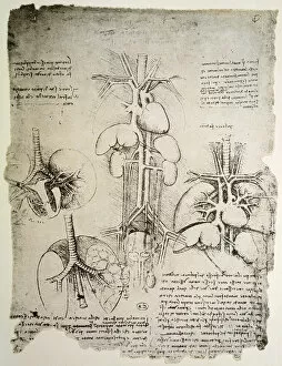
Background imageDissection Collection: Dissection of the Principal Organs of a Woman, c1480 (1945). Artist: Leonardo da Vinci

Dissection of the Principal Organs of a Woman, c1480 (1945). Artist: Leonardo da Vinci
Dissection of the Principal Organs of a Woman, c1480 (1945). From The Drawings of Leonardo da Vinci. [Reynal & Hitchcock, New York, 1945]

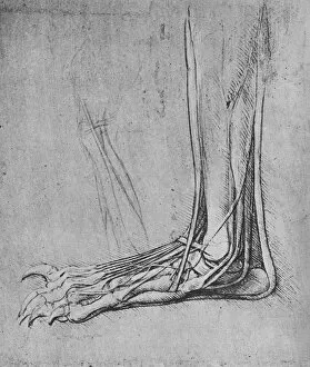
Background imageDissection Collection: Dissection of a Bears Foot to the Left, c1480 (1945). Artist: Leonardo da Vinci

Dissection of a Bears Foot to the Left, c1480 (1945). Artist: Leonardo da Vinci
Dissection of a Bears Foot to the Left, c1480 (1945). From The Drawings of Leonardo da Vinci. [Reynal & Hitchcock, New York, 1945]

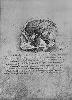
Background imageDissection Collection: Anatomical Drawing of a Skull to the Left, c1480 (1945). Artist: Leonardo da Vinci

Anatomical Drawing of a Skull to the Left, c1480 (1945). Artist: Leonardo da Vinci
Anatomical Drawing of a Skull to the Left, c1480 (1945). From The Drawings of Leonardo da Vinci. [Reynal & Hitchcock, New York, 1945]

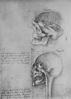
Background imageDissection Collection: Anatomical Drawings of Two Skulls in Profile to the Left, c1480 (1945). Artist: Leonardo da Vinci

Anatomical Drawings of Two Skulls in Profile to the Left, c1480 (1945). Artist: Leonardo da Vinci
Anatomical Drawings of Two Skulls in Profile to the Left, c1480 (1945). From The Drawings of Leonardo da Vinci. [Reynal & Hitchcock, New York, 1945]

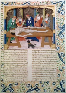
Background imageDissection Collection: Dissection, late 15th century

Dissection, late 15th century. Manuscript page from a French version of De proprietatibus rerum by Bartholomaeus Anglicus, in the collection of the Bibliotheque Nationale, Paris

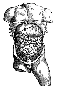
Background imageDissection Collection: Abdominal cavity and its contents, 1543

Abdominal cavity and its contents, 1543. From De humani corporis fabrica by Andreas Vesalius. (Basle, 1543)

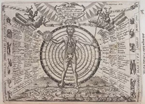
Background imageDissection Collection: An astrological chart, 1646. Artist: Athanasius Kircher

An astrological chart, 1646. Artist: Athanasius Kircher
An astrological chart, 1646. A man with his internal organs revealed standing in front of a circular chart. One hand has been replaced by a sun

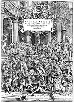
Background imageDissection Collection: Title page of Andreas Vesalius De Humani Corporis Fabrica, showing Vesalius dissecting body, 1543

Title page of Andreas Vesalius De Humani Corporis Fabrica, showing Vesalius dissecting body, 1543
Title page of Andreas Vesalius De Humani Corporis Fabrica, showing Vesalius dissecting a body, 1543

Background imageDissection Collection: Late 16th century anatomy theatre, Jacques de Gehyn the Elder, 1633

Late 16th century anatomy theatre, Jacques de Gehyn the Elder, 1633. Artist: Jacques de Gehyn the Elder
Late 16th century anatomy theatre by Jacques de Gehyn the Elder, 1633. From Dutch translation by Vopiscus Plemp of Barthelemy Cabrol Anatomical Alphabet, Amsterdam, 1633. Engraving

Background imageDissection Collection: Andrea Vesalio. De humani corporis fabrica

Andrea Vesalio. De humani corporis fabrica
Vesalius, Andreas (1514-1564). Brabantian anatomist, physician, and author of one of the most influential books on human anatomy, De humani corporis fabrica. Oil

Background imageDissection Collection: Naked male on a plinth with cross section of stomach

Naked male on a plinth with cross section of stomach
Tab II. Naked male on a plinth with cross section of stomach Source: Myographia nova, or, A graphical description of all the muscles in the humane body, as they arise in dissection by Browne, John

Background imageDissection Collection: Naked man on plinth with cross section of muscles

Naked man on plinth with cross section of muscles
Praelectio secunda - Tab I. Naked man on plinth with cross section of side and shoulder muscles Source: Myographia nova, or, A graphical description of all the muscles in the humane body

Background imageDissection Collection: Anatomy of the human skin

Anatomy of the human skin

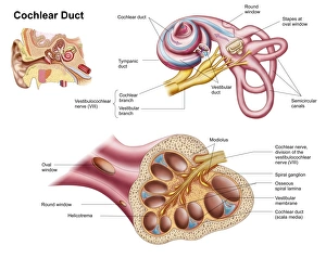
Background imageDissection Collection: Anatomy of the cochlear duct in the human ear

Anatomy of the cochlear duct in the human ear

Background imageDissection Collection: Anatomy of human ear

Anatomy of human ear

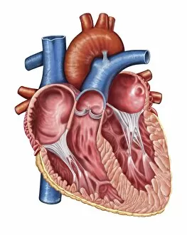
Background imageDissection Collection: Interior of human heart

Interior of human heart

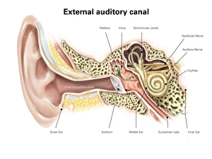
Background imageDissection Collection: External auditory canal of human ear (with labels)

External auditory canal of human ear (with labels)


All Professionally Made to Order for Quick Shipping

"Unveiling the Wonders Within: Exploring the Intricacies of Dissection" Step into a world where art and science intertwine as we delve into the captivating realm of dissection. From Leonardo da Vinci's meticulous study of skull anatomy to Rembrandt's haunting depiction of anatomical dissections, these masterpieces offer us a glimpse into the intricate workings of the human body. Witness how Leonardo da Vinci's unrivaled genius comes alive in his Skull Anatomy, revealing every contour and detail with astonishing precision. Marvel at his ability to unravel the mysteries hidden beneath our skin, showcasing not only beauty but also scientific accuracy. Venture further and explore the beating heart, an organ that symbolizes life itself. Discover its complex network of blood vessels intricately depicted by skilled artists throughout history. These illustrations serve as a testament to our relentless pursuit in understanding this vital organ. Continue your journey through time and witness groundbreaking works like De humani corporis fabrica by Andreas Vesalius, which revolutionized anatomical studies during the Renaissance period. Delve deeper into Rembrandt's Dissection series, capturing both fascination and unease surrounding this practice. Feast your eyes on detailed drawings depicting musculature from various angles; each stroke meticulously crafted to showcase every fiber and sinew. These artworks remind us that beneath our skin lies a symphony of muscles working tirelessly together. Immerse yourself in The Anatomy Lesson of Dr Nicolaes Tulp by Rembrandt Harmensz van Rijn—a masterpiece that captures both scientific inquiry and artistic brilliance. Witness how physicians gather around their subject for an educational experience unlike any other. Finally, gaze upon breathtaking artwork illustrating arteries coursing through head, chest, male groin—each stroke telling stories untold until now. These visual narratives transport us back centuries ago when exploration was paramount in unlocking medical knowledge. Join us on this extraordinary journey where art meets science—a celebration of the human body's complexity and our relentless pursuit of knowledge.