Fraud Blocker Skip to main content

Dissection Collection (page 9)

"Unveiling the Wonders Within

Background imageDissection Collection: English physician and anatomist. Harvey demonstrating his theory of the circulation of the blood

English physician and anatomist. Harvey demonstrating his theory of the circulation of the blood to King Charles I
WILLIAM HARVEY (1578-1657). English physician and anatomist. Harvey demonstrating his theory of the circulation of the blood to King Charles I and the future King Charles II

Background imageDissection Collection: Cuttlefish dissection drawing

Cuttlefish dissection drawing
Table LI, taken from Bibel der Natur byJan Swammerdamm

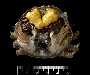
Background imageDissection Collection: Dissected female Eriocheir sinensis, Chinese mitten crab

Dissected female Eriocheir sinensis, Chinese mitten crab
A dissected female mitten crab (eriocheir sinensis), showing the ripening ovaries. Specimen was collected from the River Thames

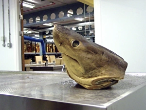
Background imageDissection Collection: Lamna nasus, porbeagle shark

Lamna nasus, porbeagle shark
Specimen of a porbeagle sharks head on the dissection table outside the Tank Room of the Darwin Centre, at the Natural History Museum, London

Background imageDissection Collection: Scientist at work at The Natural History Museum, London

Scientist at work at The Natural History Museum, London
Accurate identification of copepod crustaceans is a time consuming task for specialists, requiring meticulous sorting, dissection, and the use of high resolution microscopy

Background imageDissection Collection: ALBRECHT VON HALLER (1708-1777). Swiss scientist and poet. Haller in his laboratory at Berne

ALBRECHT VON HALLER (1708-1777). Swiss scientist and poet. Haller in his laboratory at Berne, Switzerland. Wood engraving, French, 19th century

Background imageDissection Collection: GUSTAVE FLAUBERT (1821-1880). French novelist. Caricature by Achille Lemot of Flaubert dissecting

GUSTAVE FLAUBERT (1821-1880). French novelist. Caricature by Achille Lemot of Flaubert dissecting Madame Bovary, 1869

Background imageDissection Collection: KANGAROO: ANATOMY. Dissected head of young kangaroo. Line engraving, 19th century

KANGAROO: ANATOMY. Dissected head of young kangaroo. Line engraving, 19th century

Background imageDissection Collection: RICHTER ILLUSTRATION. Wood engraving, 19th century, by August Gaber after Ludwig Richter (1803-1884)

RICHTER ILLUSTRATION. Wood engraving, 19th century, by August Gaber after Ludwig Richter (1803-1884)

Background imageDissection Collection: WHALE DISSECTION. The cutting up of a whale. Line engraving, French, 18th century

WHALE DISSECTION. The cutting up of a whale. Line engraving, French, 18th century

Background imageDissection Collection: WHALING, c1840. Whale Fishery - Cutting up a Whale. Line engraving from an American school

WHALING, c1840. Whale Fishery - Cutting up a Whale. Line engraving from an American school geography, c1840

Background imageDissection Collection: WHALING, LONG ISLAND, 1885. Cutting up a right whale caught off Southampton, Long Island, in 1885

WHALING, LONG ISLAND, 1885. Cutting up a right whale caught off Southampton, Long Island, in 1885. Wood engraving from a contemporary American newspaper

Background imageDissection Collection: Hyacinth Bulb

Hyacinth Bulb
A hyacinth bulb cut in two, with a section clearly showing the leaves and flower inside the bulb. Date: 1960s

Background imageDissection Collection: Herpetologist taking liver from frog for genetic study purposes, Manu Road, Departemento Cuzco

Herpetologist taking liver from frog for genetic study purposes, Manu Road, Departemento Cuzco, Andes, Peru

Background imageDissection Collection: Muscles of the head and neck

Muscles of the head and neck, historical anatomical artwork. This head has been dissected to show several muscles involved in moving the face and neck

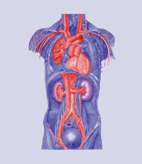
Background imageDissection Collection: Torso blood vessels

Torso blood vessels. Historical artwork of a human torso that has been dissected to show major blood vessels. The heart (upper centre) pumps blood to the lungs on either side (to be oxygenated)

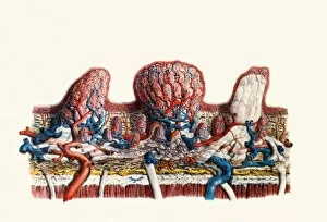
Background imageDissection Collection: Papillae on the tongue

Papillae on the tongue
Papillae on tongue, coloured historical artwork. On the tongue most tastebuds, the structures that provide information about the taste of food, sit on small platforms called papillae

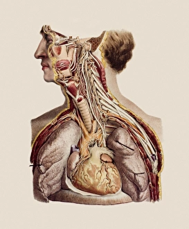
Background imageDissection Collection: Cranial nerves

Cranial nerves, historical anatomical artwork. This neck and upper torso have been dissected to show the paths of the twelve cranial nerves (white)

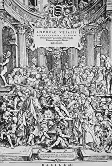
Background imageDissection Collection: Title page to Vesalius book on anatomy

Title page to Vesalius book on anatomy
Title page from De Humani Corporis Fabrica (On the Structure of the Human Body) by Andreas Vesalius (1514-1564), Flemish physician & anatomist

Background imageDissection Collection: 14th Century depiction of dissection

14th Century depiction of dissection
Human dissection, 14th Century style. This image is the earliest known representation of a dissection taking place. The female corpse (right) has had most of her internal organs removed

Background imageDissection Collection: 15th century anatomical lecture

15th century anatomical lecture
15th century woodcut of an anatomical lecture at the University of Padua, Italy, showing the professor standing in his professorial chair (background)

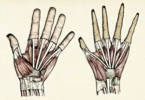
Background imageDissection Collection: Human and ape hands

Human and ape hands. Historical artwork of the hand of a human (left) and a barbary ape (Macaca sylvanus, right). The skin has been removed to reveal the underlying muscles and tendons

Background imageDissection Collection: Kidneys, nerves and blood vessels

Kidneys, nerves and blood vessels
Kidneys, historical anatomical artwork. This dorsal (back) view shows the kidneys and associated nerves lying outside the peritoneum (membrane lining the abdominal cavity)

Background imageDissection Collection: Uterus of a pregnant woman

Uterus of a pregnant woman. Artwork from William Hunters Anatomy of the Human Gravid Uterus, published 1774, showing the uterus of a woman at full-term

Background imageDissection Collection: Respiratory system

Respiratory system, anterior (front) view. Artwork taken from The Viscera of the Human Body (1840), a textbook edited by Jones Quain and Erasmus Wilson

Background imageDissection Collection: Abdominal organs

Abdominal organs, historical anatomical artwork. This ventral (front) view of a dissected abdomen shows several of the organs involved in digestion. The stomach has been removed

Background imageDissection Collection: Foot nerves

Foot nerves. Historical anatomical artwork of the nerves (white) of the sole of a human foot. Also shown are muscles (red), and ligaments, tendons and other connective tissues (grey)

Background imageDissection Collection: Leg nerves

Leg nerves. Historical anatomical artwork of the nerves (white) of the rear of a human lower leg. At left, the superficial (surface) nerves are seen and at right the deep nerves and muscles (red)

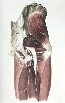
Background imageDissection Collection: Pelvic spinal nerves

Pelvic spinal nerves. Historical anatomical artwork of the spinal nerves (white) in the human pelvic region. This side view (front of the body at right)

Background imageDissection Collection: Abdominal spinal nerves

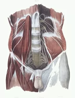
Abdominal spinal nerves. Historical anatomical artwork of the spinal nerves (white) in a human abdomen. This view, from the front

Background imageDissection Collection: Torso nerves

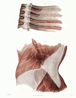
Torso nerves. Historical anatomical artwork of the nerves (white) of parts of a human torso. At lower frame, a side view of the abdomen (front of body at left) shows muscles (red)

Background imageDissection Collection: Arm nerves

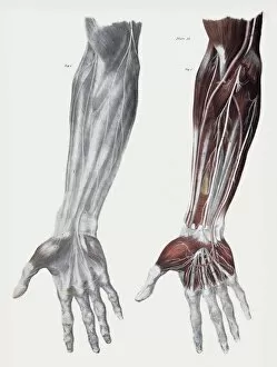
Arm nerves. Historical anatomical artwork of the nerves (white) of the front (palm side) of a human forearm. At left, the superficial (surface) nerves are shown

Background imageDissection Collection: Abdominal aorta

Abdominal aorta, historical anatomical artwork. The abdominal cavity of this torso has been dissected to give an anterior (front) view of the aorta (orange, inverted Y- shape)

Background imageDissection Collection: Knee bones and ligaments

Knee bones and ligaments. Historical anatomical artwork of knee bones (yellow) and ligaments (pale blue). Ligaments are bands of fibrous tissue that hold bones together at joints

Background imageDissection Collection: Hand bones and ligaments

Hand bones and ligaments. Historical anatomical artwork of the bones (yellow) & ligaments (white) of the hand. Ligaments are bands of fibrous tissue that hold bones together at their joints

Background imageDissection Collection: Lower arm bones and ligaments

Lower arm bones and ligaments
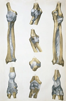
Lower arm bones & ligaments. Historical anatomical artwork of lower arm bones (yellow) and ligaments (pale blue). Ligaments are bands of fibrous tissue that hold bones together at joints

Background imageDissection Collection: Aortic aneurysm

Aortic aneurysm. Artwork from Jean Cruveilhiers medical textbook Anatomie pathalogique du corps humain, published 1835. The two large illustrations are views of an aortic aneurysm (brown, centre)

Background imageDissection Collection: Abdominal organs and nerves

Abdominal organs and nerves, historical anatomical artwork. This ventral (front) view shows an abdomen dissected to reveal some of the abdominal organs and associated nerves

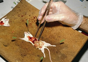
Background imageDissection Collection: Biology lesson: gloved hand dissects a mouse

Biology lesson: gloved hand dissects a mouse
Mouse dissection. Biology lesson in which a gloved hand uses a pair of forceps to dissect a white laboratory mouse Mus musculus

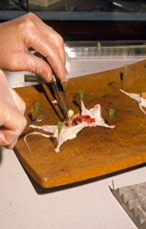
Background imageDissection Collection: Biology lesson: gloved hands dissecting a mouse

Biology lesson: gloved hands dissecting a mouse
Mouse dissection. Biology lesson in which gloved hands use intruments to dissect a white laboratory mouse Mus musculus

Background imageDissection Collection: Nerve physiology lesson, 19th century

Nerve physiology lesson, 19th century
Nerve physiology lesson. 19th-century artwork of the French physiologist Claude Bernard (1813-1878) demonstrating the role of vasomotor nerves (nerves affecting blood vessels) in a rabbit

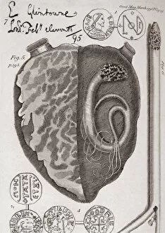
Background imageDissection Collection: Engraving of a dissected heart

Engraving of a dissected heart
18th century dissection of a heart. This engraving purports to show a serpent that was found in the left ventricle of the heart (right hand side)

Background imageDissection Collection: Dissected mouse

Dissected mouse preserved in formalin

Background imageDissection Collection: Dissected rat

Dissected rat preserved in formalin

Background imageDissection Collection: Dissected frog

Dissected frog. Common frog (Rana temporaria) dissected to show the anatomy of the blood vessels

Background imageDissection Collection: Dissected dogfish

Dissected dogfish. Preserved dogfish (Scyliorhinus canicula) dissected to show the heart and surrounding blood vessels

Background imageDissection Collection: Preserved sheeps kidney

Preserved sheeps kidney
Sheeps kindey. Dissected sheeps kidney preserved in a jar

Background imageDissection Collection: Bony labyrinth, 1844 artwork

Bony labyrinth, 1844 artwork
Bony labyrinth, external. This anatomical artwork is figure 1, plate 82 from volume 3 (1844) of Traite complet de l anatomie de l homme (1831-1854)


All Professionally Made to Order for Quick Shipping

"Unveiling the Wonders Within: Exploring the Intricacies of Dissection" Step into a world where art and science intertwine as we delve into the captivating realm of dissection. From Leonardo da Vinci's meticulous study of skull anatomy to Rembrandt's haunting depiction of anatomical dissections, these masterpieces offer us a glimpse into the intricate workings of the human body. Witness how Leonardo da Vinci's unrivaled genius comes alive in his Skull Anatomy, revealing every contour and detail with astonishing precision. Marvel at his ability to unravel the mysteries hidden beneath our skin, showcasing not only beauty but also scientific accuracy. Venture further and explore the beating heart, an organ that symbolizes life itself. Discover its complex network of blood vessels intricately depicted by skilled artists throughout history. These illustrations serve as a testament to our relentless pursuit in understanding this vital organ. Continue your journey through time and witness groundbreaking works like De humani corporis fabrica by Andreas Vesalius, which revolutionized anatomical studies during the Renaissance period. Delve deeper into Rembrandt's Dissection series, capturing both fascination and unease surrounding this practice. Feast your eyes on detailed drawings depicting musculature from various angles; each stroke meticulously crafted to showcase every fiber and sinew. These artworks remind us that beneath our skin lies a symphony of muscles working tirelessly together. Immerse yourself in The Anatomy Lesson of Dr Nicolaes Tulp by Rembrandt Harmensz van Rijn—a masterpiece that captures both scientific inquiry and artistic brilliance. Witness how physicians gather around their subject for an educational experience unlike any other. Finally, gaze upon breathtaking artwork illustrating arteries coursing through head, chest, male groin—each stroke telling stories untold until now. These visual narratives transport us back centuries ago when exploration was paramount in unlocking medical knowledge. Join us on this extraordinary journey where art meets science—a celebration of the human body's complexity and our relentless pursuit of knowledge.