Home > Popular Themes > Human Body
Muscles of the head and neck

![]()
Wall Art and Photo Gifts from Science Photo Library
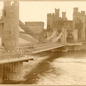
Muscles of the head and neck
Muscles of the head and neck, historical anatomical artwork. This head has been dissected to show several muscles involved in moving the face and neck. These include: The sternocleidomastoideus, running from behind the ear to the sternum, the orbicularis oris surrounding the mouth, the nasalis of the nose, and the masseter, which controls the chewing motion of the jaw. In this dissection, the square-shaped larynx (white) can also be seen partially obscured in the neck. This illustration is taken from the 19th century French textbook The Atlas of Human Anatomy and Surgery by J. M. Bourgery and N. H. Jacob
Science Photo Library features Science and Medical images including photos and illustrations
Media ID 6448505
© MEHAU KULYK/SCIENCE PHOTO LIBRARY
Atlas Of Human Anatomy Attachment Cervical Dissected Dissection Face Facial French Historical Image Imagery J M Bourgery Laryngeal Larynx Masseter Muscle System Muscles Muscular N H Jacob Neck Orbicularis Oris Origin Surgery Surgical Insert Insertion Musculature Nasalis
EDITORS COMMENTS
This print showcases a historical anatomical artwork depicting the intricate muscles of the head and neck. The dissection reveals several key muscles responsible for facial and neck movements, offering a fascinating glimpse into human anatomy. Among them are the sternocleidomastoideus, which extends from behind the ear to the sternum, and plays a crucial role in turning and tilting the head. Surrounding the mouth is the orbicularis oris muscle, contributing to various facial expressions. The nasalis muscle of the nose can also be observed in this dissection, highlighting its involvement in nasal movements. Additionally, attention is drawn towards the masseter muscle that controls jaw movement during chewing. Notably visible within this image is a partially obscured square-shaped larynx situated in the neck region. This particular illustration originates from "The Atlas of Human Anatomy and Surgery" an esteemed 19th-century French textbook authored by J. M. Bourgery and N. H. Jacob. With its rich historical significance, this artwork serves as an invaluable resource for those interested in biology, surgical procedures, or simply exploring our complex muscular system. Its detailed imagery provides insights into how these muscles originate and insert within our bodies while shedding light on their vital functions throughout history.
MADE IN AUSTRALIA
Safe Shipping with 30 Day Money Back Guarantee
FREE PERSONALISATION*
We are proud to offer a range of customisation features including Personalised Captions, Color Filters and Picture Zoom Tools
SECURE PAYMENTS
We happily accept a wide range of payment options so you can pay for the things you need in the way that is most convenient for you
* Options may vary by product and licensing agreement. Zoomed Pictures can be adjusted in the Cart.