Fraud Blocker Skip to main content

Dissection Collection

"Unveiling the Wonders Within

Background imageDissection Collection: Skull anatomy by Leonardo da Vinci

Skull anatomy by Leonardo da Vinci. Historical artwork and notes on the anatomy of the human skull and teeth, by the Italian artist and scientist Leonardo da Vinci (1452-1519)

Background imageDissection Collection: Heart

Heart. Historical anatomical artwork of the human heart, seen from the front. Coronary blood vessels are seen on the surface of the heart

Background imageDissection Collection: The Anatomy Lesson of Dr Nicolaes Tulp, 1632. Artist: Rembrandt Harmensz van Rijn

The Anatomy Lesson of Dr Nicolaes Tulp, 1632. Artist: Rembrandt Harmensz van Rijn
The Anatomy Lesson of Dr Nicolaes Tulp, 1632. Rembrandt van Rhijn (1606-1669). Found in the collection of the The Mauritshuis, The Hague

Background imageDissection Collection: Anatomy / Sole of Foot

Anatomy / Sole of Foot
First and second stages of the dissection of the sole of the foot

Background imageDissection Collection: De humani corporis fabrica (Of the Structure of the Human Body), 1555

De humani corporis fabrica (Of the Structure of the Human Body), 1555

Background imageDissection Collection: Male groin arteries, 1825 artwork

Male groin arteries, 1825 artwork
Male groin arteries. Dissections of a male groin to show areas supplied by the internal iliac artery (red) and its branches

Background imageDissection Collection: Heart and lungs

Heart and lungs. Historical anatomical artwork of the human heart and lungs, seen from the front. Dissection hooks have been used to draw back the lungs (red)

Background imageDissection Collection: Rembrandts Dissection

Rembrandts Dissection
An anatomical demonstration

Background imageDissection Collection: Anatomical drawing of musculature, side view

Anatomical drawing of musculature, side view
Secunda Musculorum Tabulae (Anatomical drawing of musculature, side view) Source: Andreae Vesalii Bruxellensis, scholae medicorum Patauinae professoris

Background imageDissection Collection: Influence of the Universe, the Macrocosm, on Man, the Microcosm, 1678

Influence of the Universe, the Macrocosm, on Man, the Microcosm, 1678. From Mundus Subterraneus by Athanasius Kircher. (Amsterdam, 1678)

Background imageDissection Collection: Head and chest arteries, 1825 artwork

Head and chest arteries, 1825 artwork
Head and chest arteries. Superficial arteries (red) of the chest, neck and head, which are visible when the skin and muscle fascia are removed

Background imageDissection Collection: Liver cirrhosis

Liver cirrhosis. Artwork taken from Sir Robert Carswells textbook Pathological Anatomy, published 1838. Cirrhosis is a disease in which the liver tissue is replaced with scar tissue

Background imageDissection Collection: Scientist working with a ragworm specimen

Scientist working with a ragworm specimen
Scientist dissecting a ragworm specimen, held at the Natural History Museum, London

Background imageDissection Collection: Facial nerves

Facial nerves. Historical anatomical artwork of a side view of a dissected human head showing the nerves (white) and muscles (red) of the face and neck

Background imageDissection Collection: Vesalius Demonstrates

Vesalius Demonstrates
Vesalius demonstrating

Background imageDissection Collection: Medical / Anatomy C16

Medical / Anatomy C16
Anatomy demonstration. Date: 16th Century

Background imageDissection Collection: Medical College for Women, New York

Medical College for Women, New York
7301631 Medical College for Women, New York.; Private Collection; (add.info.: A student dissecting a leg at the Medical College for Women on East Twelfth Street and Second Avenue, New York, USA)

Background imageDissection Collection: Dissection of the Knee Joint from the Front, Patella thrown down (colour litho)

Dissection of the Knee Joint from the Front, Patella thrown down (colour litho)
8667620 Dissection of the Knee Joint from the Front, Patella thrown down (colour litho) by English School, (20th century); Private Collection; (add.info.: Dissection of the Knee Joint from the Front)

Background imageDissection Collection: Dissection of the muscles in the hand and foot. Copperplate engraving by Robert Benard from Denis

Dissection of the muscles in the hand and foot. Copperplate engraving by Robert Benard from Denis Diderot's
FLO4611571 Dissection of the muscles in the hand and foot. Copperplate engraving by Robert Benard from Denis Diderot's Encyclopedia, Pellet, Geneva

Background imageDissection Collection: A dissection in progress. From Le livre de la propriete des choses (On the Properites of Things)

A dissection in progress. From Le livre de la propriete des choses (On the Properites of Things)
527491 A dissection in progress. From Le livre de la propriete des choses (On the Properites of Things) written by the English Franciscan monk Bartholomew Glanville called Bartolomaeus Anglicus

Background imageDissection Collection: Late 16th century anatomy theater. By Jacques de Gehyn the Elder

Late 16th century anatomy theater. By Jacques de Gehyn the Elder, 1633 (engraving from Dutch translation by Vopiscus)
530332 Late 16th century anatomy theater. By Jacques de Gehyn the Elder, 1633 (engraving from Dutch translation by Vopiscus Plemp of Barthelemy Cabrol Anatomical Alphabet)

Background imageDissection Collection: An open female cuttlefish, plate from 'Histoire naturelle: les mollusques' by Georges Louis Leclerc

An open female cuttlefish, plate from "Histoire naturelle: les mollusques" by Georges Louis Leclerc
AIS5369968 An open female cuttlefish, plate from "Histoire naturelle: les mollusques" by Georges Louis Leclerc, comte de Buffon (1707-1788) (engraving); Biblioteca de Catalunya, Barcelona

Background imageDissection Collection: A Illustration depicting a Dissection, 1559 (woodcut print)

A Illustration depicting a Dissection, 1559 (woodcut print)
1764830 A Illustration depicting a Dissection, 1559 (woodcut print); Private Collection; (add.info.: A Illustration depicting a Dissection. From 'De re Anatomica Libri XV')

Background imageDissection Collection: A compleat treatise of the muscles, as they appear in the humane body, and arise in dissection'

A compleat treatise of the muscles, as they appear in the humane body, and arise in dissection'
2626853 A compleat treatise of the muscles, as they appear in the humane body, and arise in dissection'; (add.info.: Illustration from 'A compleat treatise of the muscles)

Background imageDissection Collection: A dissection in progress. From Le livre de la propriete des choses (On the Properites of Things)

A dissection in progress. From Le livre de la propriete des choses (On the Properites of Things)
527490 A dissection in progress. From Le livre de la propriete des choses (On the Properites of Things) written by the English Franciscan monk Bartholomew Glanville, called Bartolomaeus Anglicus

Background imageDissection Collection: An anatomy lecture at Padua

An anatomy lecture at Padua
5311884 An anatomy lecture at Padua; (add.info.: Woodcut engraving of an anatomy lecture at Padua. The professor remains in his chair reading from a book)

Background imageDissection Collection: Medical College for Women, New York

Medical College for Women, New York
7301630 Medical College for Women, New York.; Private Collection; (add.info.: Women students attend a lecture in the Anatomical Lecture Room at the Medical College for Women on East Twelfth Street)

Background imageDissection Collection: Details of the anatomy of the human ear, showing cochlea, stapes, incus, malleus

Details of the anatomy of the human ear, showing cochlea, stapes, incus, malleus, semicircular canal, etc
FLO4611637 Details of the anatomy of the human ear, showing cochlea, stapes, incus, malleus, semicircular canal, etc. Copperplate engraving by Robert Benard after an illustration by Guichard Joseph

Background imageDissection Collection: Dissection of the human hand and foot to reveal muscle, ligament and bone

Dissection of the human hand and foot to reveal muscle, ligament and bone
FLO4611552 Dissection of the human hand and foot to reveal muscle, ligament and bone. Copperplate engraving by Robert Benard from Denis Diderot's Encyclopedia, Pellet, Geneva

Background imageDissection Collection: Dissection of the Hip Joint from the Front (colour litho)

Dissection of the Hip Joint from the Front (colour litho)
8667621 Dissection of the Hip Joint from the Front (colour litho) by English School, (20th century); Private Collection; (add.info.: Dissection of the Hip Joint from the Front)

Background imageDissection Collection: BARTHOLIN, Thomas (1616-1680). Danish physician, mathematician, and theologian

BARTHOLIN, Thomas (1616-1680). Danish physician, mathematician, and theologian
CUL4875606 BARTHOLIN, Thomas (1616-1680). Danish physician, mathematician, and theologian. He is best known for his work in the discovery of the lymphatic system in humans

Background imageDissection Collection: Title Page of Anatome Corporis Humani, 1589 (engraving)

Title Page of Anatome Corporis Humani, 1589 (engraving)
CUL4937064 Title Page of Anatome Corporis Humani, 1589 (engraving); (add.info.: VALVERDE DE AMUSCO, Juan (1525-1588). Anatome Corporis Humani. 1589. Edition printed in Venice Barcelona

Background imageDissection Collection: Dissection of animal infected with rabies. 19th century (engraving)

Dissection of animal infected with rabies. 19th century (engraving)
672843 Dissection of animal infected with rabies. 19th century (engraving); (add.info.: Rabies (From the Latin: rabies, "madness")

Background imageDissection Collection: Depiction of Dissection. 19th century (engraving)

Depiction of Dissection. 19th century (engraving)
672828 Depiction of Dissection. 19th century (engraving); Universal History Archive/UIG.

Background imageDissection Collection: The Shepherds' Calendar, 1480-90 (gold ink & ink on parchment)

The Shepherds' Calendar, 1480-90 (gold ink & ink on parchment)
8682933 The Shepherds' Calendar, 1480-90 (gold ink & ink on parchment) by French School, (15th century); Fitzwilliam Museum, University of Cambridge

Background imageDissection Collection: View of the dissection room in the Anatomical theatre of Clamart, built in 1833 - Hauts de Seine

View of the dissection room in the Anatomical theatre of Clamart, built in 1833 - Hauts de Seine
ELD4840683 View of the dissection room in the Anatomical theatre of Clamart, built in 1833 - Hauts de Seine - (View of the dissection room in the Anatomical theatre of Clamart, near Paris)

Background imageDissection Collection: The Reward of Cruelty, 1750. Creator: John Bell

The Reward of Cruelty, 1750. Creator: John Bell
The Reward of Cruelty, 1750

Background imageDissection Collection: Anatomy of heart interior, frontal section

Anatomy of heart interior, frontal section

Background imageDissection Collection: Drawing of Two Halves of a Skull, c1480 (1945). Artist: Leonardo da Vinci

Drawing of Two Halves of a Skull, c1480 (1945). Artist: Leonardo da Vinci
Drawing of Two Halves of a Skull, c1480 (1945). From The Drawings of Leonardo da Vinci. [Reynal & Hitchcock, New York, 1945]

Background imageDissection Collection: Anatomy of adrenal gland, cross section

Anatomy of adrenal gland, cross section

Background imageDissection Collection: VESALIUS/TRADE CARD

VESALIUS/TRADE CARD
ANDREAS VESALIUS Belgian anatomist, active in Italy Date: 1514 - 1564

Background imageDissection Collection: Une Fin A l Ecole Pratique, 1902 (litho)

Une Fin A l Ecole Pratique, 1902 (litho)
2562846 Une Fin A l Ecole Pratique, 1902 (litho) by Bellanger, Camille Felix (1853-1923); Musee Orfila, Paris, France; © Archives Charmet

Background imageDissection Collection: The four stages of cruelty (engraving)

The four stages of cruelty (engraving)
666300 The four stages of cruelty (engraving) by Hogarth, William (1697-1764) (after); Private Collection; (add.info.: The four stages of cruelty. The reward of cruelty)

Background imageDissection Collection: Riot at Lyon, 27th November 1768 (ink & wash on paper) (b / w photo)

Riot at Lyon, 27th November 1768 (ink & wash on paper) (b / w photo)
CHT213091 Riot at Lyon, 27th November 1768 (ink & wash on paper) (b/w photo) by French School, (18th century); Private Collection; (add.info)

Background imageDissection Collection: The Anatomy Lesson of Doctor Velpeau (1795-1867) (charcoal on paper laid on canvas)

The Anatomy Lesson of Doctor Velpeau (1795-1867) (charcoal on paper laid on canvas)
XIR71731 The Anatomy Lesson of Doctor Velpeau (1795-1867) (charcoal on paper laid on canvas) by Feyen-Perrin, Francois Nicolas Augustin (1826-88); 182x243 cm; Musee de l Assistance Publique

Background imageDissection Collection: Taxidermy (litho)

Taxidermy (litho)
7183331 Taxidermy (litho) by English School, (20th century); Private Collection; (add.info.: Taxidermy. Illustration for Encyclopedia of Sports, Games and Pastimes (Fleetway)


All Professionally Made to Order for Quick Shipping

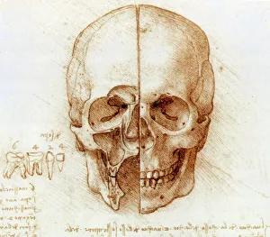
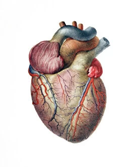
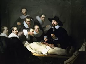
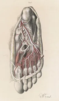
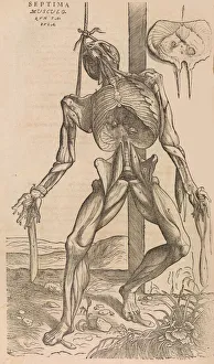
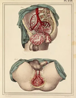
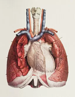
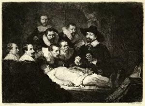
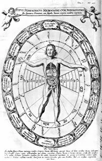
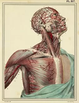
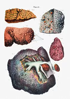
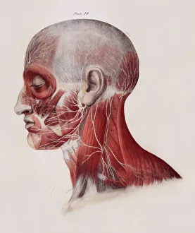
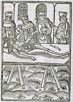
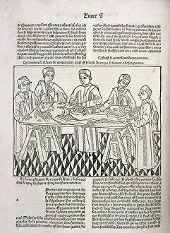
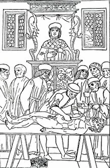
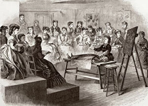
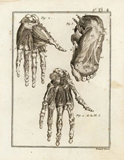
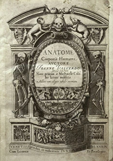
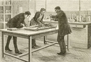
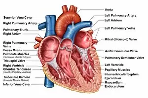
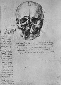
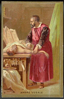
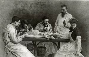
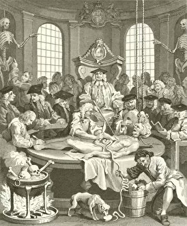
"Unveiling the Wonders Within: Exploring the Intricacies of Dissection" Step into a world where art and science intertwine as we delve into the captivating realm of dissection. From Leonardo da Vinci's meticulous study of skull anatomy to Rembrandt's haunting depiction of anatomical dissections, these masterpieces offer us a glimpse into the intricate workings of the human body. Witness how Leonardo da Vinci's unrivaled genius comes alive in his Skull Anatomy, revealing every contour and detail with astonishing precision. Marvel at his ability to unravel the mysteries hidden beneath our skin, showcasing not only beauty but also scientific accuracy. Venture further and explore the beating heart, an organ that symbolizes life itself. Discover its complex network of blood vessels intricately depicted by skilled artists throughout history. These illustrations serve as a testament to our relentless pursuit in understanding this vital organ. Continue your journey through time and witness groundbreaking works like De humani corporis fabrica by Andreas Vesalius, which revolutionized anatomical studies during the Renaissance period. Delve deeper into Rembrandt's Dissection series, capturing both fascination and unease surrounding this practice. Feast your eyes on detailed drawings depicting musculature from various angles; each stroke meticulously crafted to showcase every fiber and sinew. These artworks remind us that beneath our skin lies a symphony of muscles working tirelessly together. Immerse yourself in The Anatomy Lesson of Dr Nicolaes Tulp by Rembrandt Harmensz van Rijn—a masterpiece that captures both scientific inquiry and artistic brilliance. Witness how physicians gather around their subject for an educational experience unlike any other. Finally, gaze upon breathtaking artwork illustrating arteries coursing through head, chest, male groin—each stroke telling stories untold until now. These visual narratives transport us back centuries ago when exploration was paramount in unlocking medical knowledge. Join us on this extraordinary journey where art meets science—a celebration of the human body's complexity and our relentless pursuit of knowledge.