Home > Popular Themes > Human Body
Head and chest arteries, 1825 artwork

![]()
Wall Art and Photo Gifts from Science Photo Library
Head and chest arteries, 1825 artwork
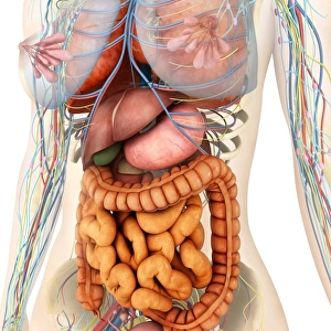
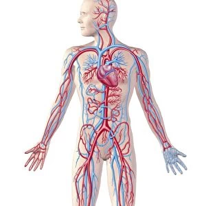
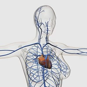
Head and chest arteries. Superficial arteries (red) of the chest, neck and head, which are visible when the skin and muscle fascia are removed. The scalp of the head has a large number of blood vessels. In the chest, the major blood vessels are located deeper in the body, below layers of muscle. This anatomical artwork is plate 217 from volume 4 of Manuel d anatomie descriptive du corps humain (1825). This 5-volume anatomy atlas was produced by French physician and surgeon Jules Germain Cloquet (1790-1883). The illustrations were by Haincelin. Volume 4 illustrated the anatomy of the circulatory and respiratory systems
Science Photo Library features Science and Medical images including photos and illustrations
Media ID 9223369
© SCIENCE PHOTO LIBRARY
1825 Anatomical Artwork Anatomical Illustration Anatomy Atlas Anterior Arterial System Arteries Blood Vessels Chest Dissected Dissection French Frontal Haincelin Jules Germain Cloquet Muscles Neck Oxygenated Blood Pectoral Scalp Superficial Thoracic Vascular Volume 4 Volume Iv Artery Blood Vessel Circulatory System
EDITORS COMMENTS
This print showcases the intricate network of head and chest arteries, providing a glimpse into the inner workings of our circulatory system. Created in 1825 by French physician and surgeon Jules Germain Cloquet, this artwork is part of his renowned five-volume anatomy atlas titled "Manuel d'anatomie descriptive du corps humain". The image reveals the superficial arteries that course through the chest, neck, and head when the skin and muscle fascia are removed. The scalp of the head is particularly rich in blood vessels while major blood vessels in the chest lie deeper beneath layers of muscle. With its historical significance and meticulous detail, this anatomical illustration offers valuable insights into human anatomy during the 19th century. It highlights how oxygenated blood flows through these vital pathways to nourish various organs and tissues. The collaboration between Cloquet and illustrator Haincelin brings this scientific knowledge to life with precision and artistry. Volume 4 specifically focuses on dissecting both circulatory and respiratory systems, making it an invaluable resource for medical professionals studying human physiology. As we gaze upon this remarkable piece, we are reminded of our body's complexity and resilience. It serves as a testament to mankind's continuous exploration of medicine throughout history. This print from Science Photo Library allows us to appreciate not only its educational value but also its aesthetic appeal as a work of art capturing our fascination with human biology.
MADE IN AUSTRALIA
Safe Shipping with 30 Day Money Back Guarantee
FREE PERSONALISATION*
We are proud to offer a range of customisation features including Personalised Captions, Color Filters and Picture Zoom Tools
SECURE PAYMENTS
We happily accept a wide range of payment options so you can pay for the things you need in the way that is most convenient for you
* Options may vary by product and licensing agreement. Zoomed Pictures can be adjusted in the Cart.