Home > Arts > Street art graffiti > Digital art > Digital paintings
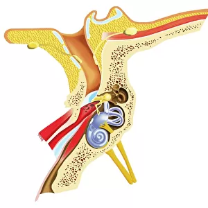
External auditory canal of human ear (with labels)
")
![]()
Wall Art and Photo Gifts from Stocktrek
External auditory canal of human ear (with labels)
Stocktrek Images specializes in Astronomy, Dinosaurs, Medical, Military Forces, Ocean Life, & Sci-Fi
Media ID 13011119
© Alan Gesek/Stocktrek Images
Acoustic Anatomy Audio Auditory Auditory System Aural Auricle Biology Biomedical Illustrations Cochlea Cross Section Cutaway View Detail Diagram Dissection Ear Canal Ear Drum Eustachian Tube Healthcare Healthy Hearing Human Anatomy Human Body Human Body Parts Human Ear Human Organs Human Tissue Incus Inner Ear Internal Organs Listening Lobe Malleus Meatus Medical Medicine Membrane Middle Ear Nerve Organ Organ Of Corti Ossicle Outer Ear Physiology Pinna Scarpas Ganglion Sensory System Sound Stapes Structure Tympanic Membrane Utricle Vestibular System Labyrinth
FEATURES IN THESE COLLECTIONS
> Arts
> Street art graffiti
> Digital art
> Digital paintings
> Posters
> Aircraft Posters
> Cutaway Posters
EDITORS COMMENTS
This print showcases the intricate beauty of the external auditory canal in the human ear. With meticulous labeling, this horizontal, digitally generated image provides a comprehensive understanding of the auditory system and its various components. From the eustachian tube to the labyrinth, every detail is meticulously depicted on a clean white background. The dissection-like cutaway view allows us to explore deeper into this vital organ responsible for our sense of hearing. The pinna, or outer ear, leads us into the ear canal where we encounter the tympanic membrane or eardrum. Moving further inward, we discover delicate structures such as ossicles - malleus, incus, and stapes - which transmit sound vibrations to the inner ear. The cochlea takes center stage within this artwork; it is here that sound waves are transformed into electrical signals that can be interpreted by our brain. Alongside it lies another crucial component called Scarpas ganglion which plays a significant role in transmitting sensory information. This visually stunning illustration not only serves as an educational tool but also highlights the remarkable complexity and functionality of our auditory system. It invites us to marvel at how these internal organs work harmoniously to enable one of our most precious senses – hearing. Alan Gesek's expertise shines through in this biomedical masterpiece that seamlessly blends science with artistry.
MADE IN AUSTRALIA
Safe Shipping with 30 Day Money Back Guarantee
FREE PERSONALISATION*
We are proud to offer a range of customisation features including Personalised Captions, Color Filters and Picture Zoom Tools
SECURE PAYMENTS
We happily accept a wide range of payment options so you can pay for the things you need in the way that is most convenient for you
* Options may vary by product and licensing agreement. Zoomed Pictures can be adjusted in the Cart.