
Pinna Collection
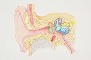
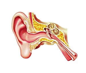
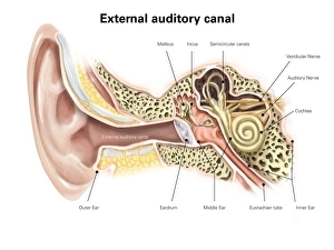
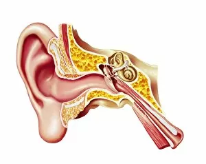
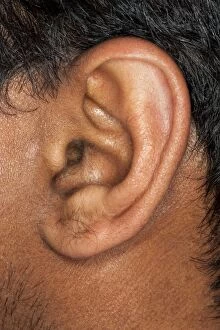
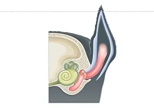
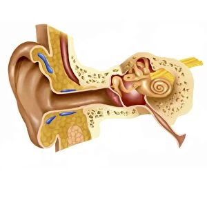
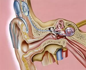
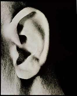
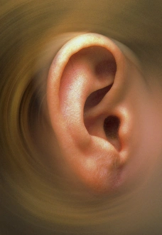
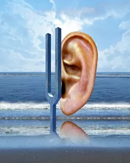
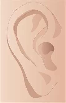
The pinna, also known as the auricle, is an essential part of the human ear and can be visualized in a cross-section diagram of the ear


All Professionally Made to Order for Quick Shipping
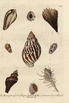
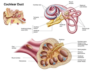
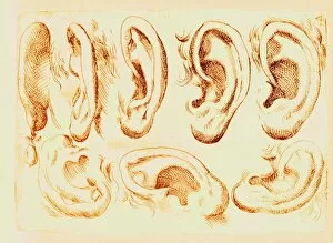
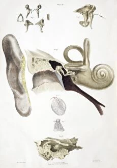
The pinna, also known as the auricle, is an essential part of the human ear and can be visualized in a cross-section diagram of the ear, showcasing its unique structure and function. Just like shells found in creatures such as clams, mussels, oysters, scallops, and more, the pinna serves as a protective covering for our delicate auditory system. While these marine creatures have their own remarkable shells to safeguard them from external elements, humans rely on their pinna to collect sound waves and direct them into the external auditory canal. This fascinating organ allows us to perceive sounds with incredible precision. Speaking of shells resembling our pinna's shape, one notable example is the noble pen shell (Pinna nobilis), which happens to be the largest bivalve species in the Mediterranean region. Its magnificent shell bears resemblance to our very own auricle. Another intriguing member of this family is the baggy pen shell or bag pinna (Pinna saccata). With its distinct appearance and intricate design patterns on its shell surface, it reminds us of both nature's creativity and our own anatomical wonders. Various specimens of shells further emphasize how diverse shapes can be found within this group. Each specimen showcases nature's ability to create stunning structures that serve different purposes – just like how each individual's unique pinna contributes to their hearing experience. Delving deeper into human anatomy reveals detailed diagrams illustrating not only outer structures but also inner components associated with hearing mechanisms. The cochlear duct plays a crucial role in converting sound vibrations into electrical signals that are then transmitted through nerves for interpretation by our brain. The external auditory canal labeled within an illustration provides insight into how sound travels from outside towards our eardrum – a journey facilitated by none other than our trusty pinna. Historical engravings from sources like The Pictorial Museum of Animated Nature capture early attempts at depicting various aspects related to the human ear.