Home > Arts > Still life artwork > Watercolor paintings > Fine art
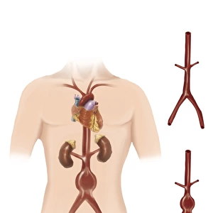
Interior detail of the cochlea

![]()
Wall Art and Photo Gifts from Stocktrek
Interior detail of the cochlea
Stocktrek Images specializes in Astronomy, Dinosaurs, Medical, Military Forces, Ocean Life, & Sci-Fi
Media ID 13013379
© TriFocal Communications/Stocktrek Images
Acoustic Anatomy Audio Auditory Auditory System Aural Auricle Basilar Membrane Biology Biomedical Illustrations Cell Cochlea Cochlear Duct Cross Section Cutaway View Detail Digitally Enhanced Dissection Ear Canal Ear Drum Endolymph Gland Health Healthcare Hearing Human Anatomy Human Body Human Body Parts Human Ear Human Glands Human Organs Human Tissue Illustration Technique Incus Inner Ear Internal Organs Listening Magnification Malleus Meatus Medical Medicine Membrane Membranous Labyrinth Microvillus Middle Ear Nerve Organ Organ Of Corti Organelle Ossicle Outer Ear Physiology Pinna Scala Tympani Scala Vestibuli Scarpas Ganglion Sensory Receptors Sensory System Sound Spiral Ganglion Stapes Structure Tympanic Membrane Utricle Vestibular Nerves Vestibular System Watercolor Painting Hair Cells Modiolus Semicircular Canal Stereocilia
FEATURES IN THESE COLLECTIONS
> Arts
> Still life artwork
> Watercolor paintings
> Fine art
> Arts
> Street art graffiti
> Digital art
> Digital paintings
> Arts
> Watercolor paintings
> Watercolor illustrations
> Posters
> Aircraft Posters
> Cutaway Posters
EDITORS COMMENTS
This watercolor painting, digitally enhanced to perfection, showcases the intricate interior detail of the cochlea. With its vibrant colors and impeccable illustration technique, this artwork is a true masterpiece in biomedical illustrations. Against a crisp white background, the cross-sectioned view of the cochlea reveals its fascinating components and functions. From sensory receptors to hair cells, every tiny detail is meticulously depicted in this stunning piece. The auditory system comes alive as we explore the delicate membrane structures such as the basilar membrane and stereocilia. Magnification allows us to appreciate even the smallest organelles like microvilli within this complex organ. The vestibulocochlear nerves gracefully intertwine with other internal organs like incus, malleus, meatus, pinna, and stapes. This scientific artwork not only portrays human anatomy but also highlights how sound travels through our ears for listening pleasure. As we delve deeper into this dissection-like portrayal of the cochlea's scala tympani and scala vestibuli, our understanding of its functionality expands further. The osseous spiral lamina leads us to discover helicotrema while spiral ganglion and modiolus add depth to this mesmerizing composition. With an emphasis on human glands like endolymph and Reissner's membrane along with Scarpas ganglion and semicircular canal from the vestibular system - it becomes evident that this illustration encompasses various aspects of our sensory system. TriFocal Communications has truly captured both artistry and science in one frame with their exceptional attention to detail in portraying these essential elements of human hearing physiology.
MADE IN AUSTRALIA
Safe Shipping with 30 Day Money Back Guarantee
FREE PERSONALISATION*
We are proud to offer a range of customisation features including Personalised Captions, Color Filters and Picture Zoom Tools
SECURE PAYMENTS
We happily accept a wide range of payment options so you can pay for the things you need in the way that is most convenient for you
* Options may vary by product and licensing agreement. Zoomed Pictures can be adjusted in the Cart.