Fraud Blocker Skip to main content

Home > Popular Themes > Human Body

Ear anatomy

 : Ear anatomy
In Stock
We accept all major payment options so you can pay for the things you need in the way that is most convenient for you

Choose Your Product

Framed Photos
Buy from AU$119.99
Photo Prints
Buy from AU$12.99
Jigsaw Puzzles
Buy from AU$89.99
Poster Prints
Buy from AU$24.99
Canvas Prints
Buy from AU$124.99
Fine Art Prints
Buy from AU$92.53
Metal Prints
Buy from AU$43.99
Cards
Buy from AU$12.99
Home Decor
Buy from AU$24.99

Science Photo Library Photo Prints and Wall Art

Ear anatomy

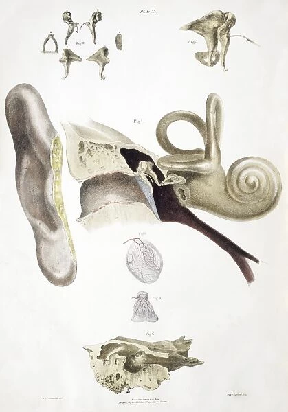
Ear anatomy. Historical anatomical artwork of a human ear. The main diagram (centre) shows the outer ear (pinna, left) and the internal structure of the ear (moving left-right): the ear canal, the ear drum (blue), the ear bones, the cochlea (snail shaped). Just before the cochlea are two other stuctures: the semicircular canals (three loops at right angles to each other), and the Eustachian tube (brown, connecting to the throat). The ear bones are also shown at top. The ear drum and its blood vessels is also at lower centre with another ear membrane below. The temporal bone (bottom) houses the ear. Artwork from The Nerves of the Human Body (Ed. Jones Quain, London, 1839). Science Photo Library features Science and Medical images including photos and illustrations. © SHEILA TERRY/SCIENCE PHOTO LIBRARY

Media ID 6448091

1839 Anvil Bones Book Canal Canals Cochlea Drawing Drum Ear Drum Hammer Jones Quain Lateral Pinna Profile Sense Side Text Book Tympanic Membrane Vestibule Eustachian Tube Sectioned Semi Circular Temporal Bone Vestibular

FEATURES IN THESE COLLECTIONS

> Animals > Insects > Snails > Related Images

> Popular Themes > Human Body

SECURE PAYMENTS
We accept all major payment options so you can pay for the things you need in the way that is most convenient for you

Made in Australia
Safe Shipping, Easy Returns

FREE PERSONALISATION
We are proud to offer a range of customisation including Cropping, Captions, and Filters. Zoom Cropping can be adjusted in the basket


EDITORS COMMENTS

This print showcases a historical anatomical artwork of the intricate ear anatomy. Dating back to 1839, this illustration from "The Nerves of the Human Body" provides a detailed depiction of the human ear's internal structure and outer features. At its center, we observe the pinna, or outer ear, on the left side while moving towards the right reveals an array of essential components. The main diagram highlights various elements including the ear canal, represented by a pathway leading inward. Just beyond lies the mesmerizing blue-colored eardrum, followed by delicate ear bones and finally culminating in the snail-shaped cochlea. Adjacent to this remarkable structure are two additional formations: three semicircular canals arranged at right angles to each other and a brown Eustachian tube connecting to the throat. Atop this comprehensive illustration are showcased both upper and lower views of the ear bones along with another membrane situated below. The temporal bone is prominently displayed at bottom as it houses this marvelously complex organ responsible for our sense of hearing. This awe-inspiring artwork not only serves as an invaluable resource for medical professionals but also offers us a glimpse into history's exploration of human anatomy. With meticulous attention to detail and artistic finesse, it stands as a testament to mankind's ongoing quest for knowledge about our own bodies.


Framed Prints of Ear anatomy

Discover the intricacies of the human ear with our captivating Framed Prints from Media Storehouse, featuring this historic anatomical artwork by Science Photo Library. This exquisite image offers a mesmerizing glimpse into the ear's complex structure, illustrating both the outer ear and the intricate inner workings, including the ear canal. Bring the wonders of science into your home or office with this striking and educational addition to your décor.


Photo Prints of Ear anatomy

Discover the intricacies of the human ear with our Media Storehouse Photographic Prints featuring this captivating image from Science Photo Library. This historical anatomical artwork brings to life the complex structure of the ear, revealing the outer ear (pinna) and the intricate inner workings, including the ear canal. Add this enlightening print to your home or office decor and ignite curiosity and conversation. The high-quality print is sure to impress and make a beautiful addition to any space.


Poster Prints of Ear anatomy

Discover the intricacies of the human ear with our captivating Ear Anatomy Poster Print from Media Storehouse, in partnership with Science Photo Library. This beautifully detailed historical artwork illustrates the outer ear and the intricate inner structure, including the ear canal. Bring the wonders of anatomy into your home or classroom with this high-quality, vibrant poster print. Perfect for educators, students, or anyone with a curiosity for the marvels of the human body. Order yours today and ignite a passion for learning!


Jigsaw Puzzles of Ear anatomy

Explore the intricacies of human anatomy with our captivating Ear Anatomy jigsaw puzzle from Media Storehouse. This puzzle, featuring an exquisite illustration from Science Photo Library, invites you on a fascinating journey into the complex world of the ear. Delve into the intricate details of the outer ear and the inner structure, including the ear canal. Ideal for anatomists, medical professionals, or anyone with a curiosity for the wonders of the human body, this educational puzzle promises a challenging yet rewarding experience. Piece together the historical artwork and unravel the mysteries of the ear, one puzzle piece at a time. Engage your mind, stimulate your creativity, and expand your knowledge with Media Storehouse's Ear Anatomy puzzle.

diamond
Made With High Grade Materials
thumb_up
Professional Quality Finish
package_2
Carefully Packed To Aid Safe Arrival

You May Also Like


Jon Bon Jovi performing at Ibrox Stadium, Glasgow, Scotland during the group Bon JoviJon Bon Jovi performing at Ibrox Stadium, Glasgow, Scotland during the group Bon Jovi
Add to Basket
King Henry V weddingKing Henry V wedding
Add to Basket
The Cathedral Saint Sauveur (b / w photo)The Cathedral Saint Sauveur (b / w photo)
Add to Basket
A Doctors Prescription (woodcut) (b / w photo)A Doctors Prescription (woodcut) (b / w photo)
Add to Basket
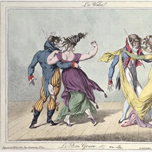
Le Bon Genre: Le Walse, after Carle Vernet (1758-1836), 1810 (hand-coloured etching)Le Bon Genre: Le Walse, after Carle Vernet (1758-1836), 1810 (hand-coloured etching)
Add to Basket
Destruction de l armee de Sennacherib (engraving)Destruction de l armee de Sennacherib (engraving)
Add to Basket
British New Year card (colour litho)British New Year card (colour litho)
Add to Basket
Wooden Cabinet Doors, c. 1939. Creator: Rose Campbell-GerkeWooden Cabinet Doors, c. 1939. Creator: Rose Campbell-Gerke
Add to Basket
Longtail boats on Phra Nang Beach at sunset, Railay Peninsula, Krabi Provonce, ThailandLongtail boats on Phra Nang Beach at sunset, Railay Peninsula, Krabi Provonce, Thailand
Add to Basket
Mutual Aid in Air Defence. The first trial in the Midland Civil Defence Region ofMutual Aid in Air Defence. The first trial in the Midland Civil Defence Region of
Add to Basket
Jon Bon Jovi performing at Ibrox Stadium, Glasgow, Scotland during the group Bon JoviJon Bon Jovi performing at Ibrox Stadium, Glasgow, Scotland during the group Bon Jovi
Add to Basket
Young woman in summer dress, 17th Century (coloured engraving)Young woman in summer dress, 17th Century (coloured engraving)
Add to Basket

Gallery Wall Inspiration

Sensory homunculus
Sensory homunculus
Plague doctor, 17th century artwork
Brain blood vessels, 3D angiogram C007 / 1981
Brain pathways
Cerebellum tissue, light micrograph
Brain fibres, DTI MRI scan C017 / 7035
Purkinje nerve cells in the cerebellum
Medulla oblongata in the brain, artwork
Nerve and glial cells, light micrograph
Motor and sensory homunculi
Nerve cell, SEM
Phrenological model of personality traits
Diagram of the human brain and spinal column
Phrenological Head / Scrap
Nerve cell
Moth antenna, SEM
Touch receptors
Hip replacement, artwork
The Sacred Heart; painting by Pompeo Batoni. Chiesa del Ges, Church of Jesus, Rome
30587745
Motor homunculus model
The Return of the Prodigal Son, c1668. Artist: Rembrandt Harmensz van Rijn
Brain fibres, DTI MRI scan C017 / 7099
Motor homunculus
Normal knees, X-ray
Human Anatomy - Backbone including Ribs and Pelvis


© Copyright Media Storehouse, All Rights Reserved 2003 - 2025
Facebook     Pinterest     100% Payment Secure     Reviews IO