Fraud Blocker Skip to main content

False Coloured Collection (page 2)

"Revealing the Unseen: Exploring the World Through False Colors" Step back in time to medieval alchemy

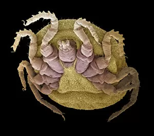
Background imageFalse Coloured Collection: Sheep tick, SEM

Sheep tick, SEM
Sheep tick. Coloured scanning electron micrograph (SEM) of a sheep tick (Ixodes ricinus). The ticks mouthparts are between its two front legs

Background imageFalse Coloured Collection: Small intestine, SEM

Small intestine, SEM
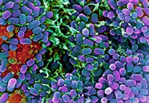
Small intestine. Coloured scanning electron micrograph (SEM) of a biopsy from the small intestines of a child. The small intestine runs from the stomach to the large intestine

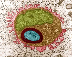
Background imageFalse Coloured Collection: Regenerating nerve cell, TEM

Regenerating nerve cell, TEM
Regenerating nerve cell. Coloured transmission electron micrograph (TEM) of a section through a nerve axon (blue) regenerating within a Schwann cell (light brown)

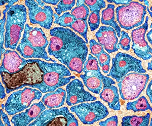
Background imageFalse Coloured Collection: Myelination of nerve fibres, TEM

Myelination of nerve fibres, TEM
Myelination of nerve fibres. Coloured transmission electron micrograph (TEM) of Schwann cells (blue, with brown nuclei) insulating nerve fibres (axons, pink) with a myelin sheath

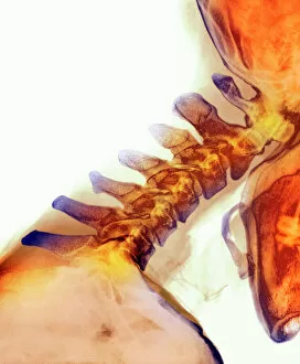
Background imageFalse Coloured Collection: Neck vertebrae extended, X-ray

Neck vertebrae extended, X-ray
Bending of the neck. Coloured X-ray of a side view of the neck of a man showing extension of healthy cervical vertebrae (bones)

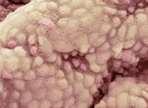
Background imageFalse Coloured Collection: Gallbladder surface, SEM

Gallbladder surface, SEM
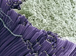
Gallbladder. Coloured scanning electron micrograph (SEM) of the surface of the gallbladder showing the numerous microvilli that cover the columnar epithelium

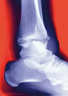
Background imageFalse Coloured Collection: Fractured ankle, X-ray

Fractured ankle, X-ray
Fractured ankle. Coloured profile X-ray of a distal fibula fracture (upper centre). The fibula is the smaller leg bone running down from top centre. The larger bone is the tibia

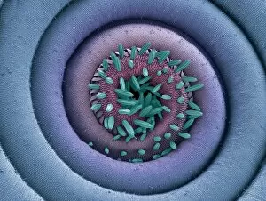
Background imageFalse Coloured Collection: Moth proboscis, SEM

Moth proboscis, SEM
Moth proboscis. Coloured scanning electron micrograph (SEM) of the coiled proboscis of a moth (order Lepidoptera). The proboscis is an elongated part of the mouth

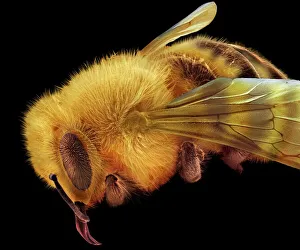
Background imageFalse Coloured Collection: Honey bee, SEM

Honey bee, SEM
Honey bee (Apis mellifera), coloured scanning electron micrograph (SEM). The head and one of the two compound eyes is at lower left

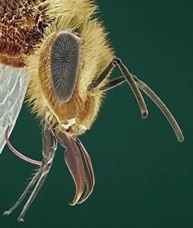
Background imageFalse Coloured Collection: Head of a honey bee, SEM

Head of a honey bee, SEM
Head of a honey bee (Apis mellifera), coloured scanning electron micrograph (SEM). The compound eyes (one seen) are either side of the antennae

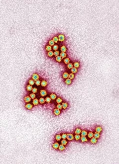
Background imageFalse Coloured Collection: Norovirus particles, TEM

Norovirus particles, TEM
Norovirus particles. Coloured transmission electron micrograph (TEM) of norovirus particles. Norovirus is a genus of RNA (ribonucleic acid)

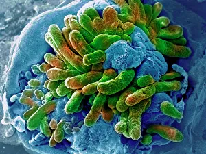
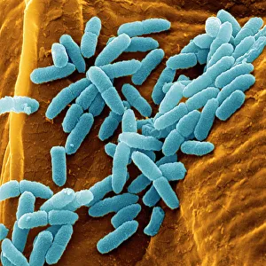
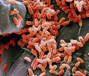
Background imageFalse Coloured Collection: Pseudomonas aeruginosa bacteria, SEM

Pseudomonas aeruginosa bacteria, SEM
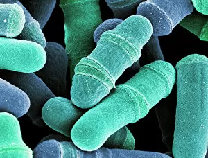
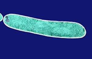
Pseudomonas aeruginosa bacteria, coloured scanning electron micrograph (SEM). These Gram-negative rod-shaped bacteria are found in soil and water, and as normal flora in the human intestine

Background imageFalse Coloured Collection: Dividing yeast cells, SEM

Dividing yeast cells, SEM
Dividing yeast cells. Coloured scanning electron micrograph (SEM) of Schizosaccharomyces pombe yeast cells dividing. S. pombe is a single-celled fungus that is studied widely as a model organism for

Background imageFalse Coloured Collection: Dividing yeast cells, SEM

Dividing yeast cells, SEM
Dividing yeast cells. Coloured scanning electron micrograph (SEM) of Schizosaccharomyces pombe yeast cells dividing. S. pombe is a single-celled fungus that is studied widely as a model organism for

Background imageFalse Coloured Collection: Bacteria infecting a macrophage, SEM

Bacteria infecting a macrophage, SEM
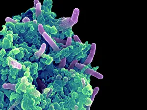
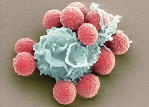
Bacteria infecting a macrophage. Coloured scanning electron micrograph (SEM) of Mycobacterium tuberculosis bacteria (purple) infecting a macrophage white blood cell

Background imageFalse Coloured Collection: E. coli bacteria, SEM

E. coli bacteria, SEM
E. coli bacteria. Coloured scanning electron micrograph (SEM) of Escherichia coli bacteria (purple) taken from the small intestine of a child. E

Background imageFalse Coloured Collection: Fat cells, TEM

Fat cells, TEM
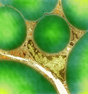
Fat cells. Coloured transmission electron micrograph (TEM) of a section through fat (adipose) tissue, consisting of fat-storing cells (adipocytes, green) supported by connective tissue

Background imageFalse Coloured Collection: Basophil white blood cell

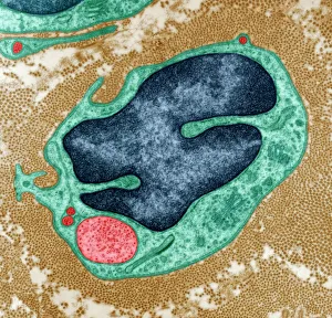
Basophil white blood cell. Coloured transmission electron micrograph (TEM) of a basophil white blood cell. Basophils are the smallest and rarest of the white blood cells

Background imageFalse Coloured Collection: False-colour photo of total solar eclipse, 1979

False-colour photo of total solar eclipse, 1979
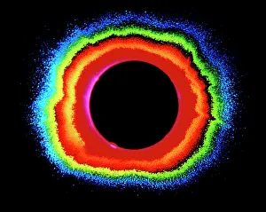
False-colour photograph of the total solar eclipse of February 26, 1979. The colour has been added by a photographic technique called isophote mapping

Background imageFalse Coloured Collection: Regenerating nerve cell, TEM

Regenerating nerve cell, TEM
Regenerating nerve cell. Coloured transmission electron micrograph (TEM) of a section through a nerve axon (yellow) regenerating within a Schwann cell (blue). The Schwann cells nucleus is black

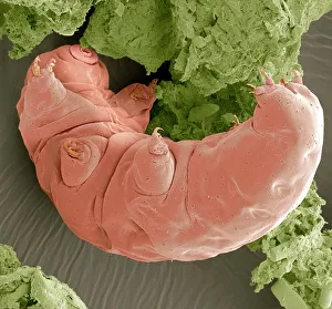
Background imageFalse Coloured Collection: Water bear, SEM

Water bear, SEM
Water bear. Coloured scanning electron micrograph (SEM) of a water bear (Macrobiotus sp.), or tardigrade. This tiny invertebrate lives in aquatic and semi-aquatic habitats such as lichen

Background imageFalse Coloured Collection: Stem cells, SEM

Stem cells, SEM
Stem cells, coloured scanning electron micrograph (SEM). Stem cells can differentiate into any other cell type. There are three main types of mammalian stem cell: embryonic stem cells

Background imageFalse Coloured Collection: Salmonella bacteria, SEM

Salmonella bacteria, SEM
Salmonella bacteria, coloured scanning electron micrograph (SEM). Each rod-shaped structure is an individual bacterium. These bacteria occur mainly in human and animal intestines

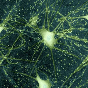
Background imageFalse Coloured Collection: Motor neurons, light micrograph

Motor neurons, light micrograph. Motor neurons are responsible for passing information around the central nervous system (CNS) and from the CNS to the rest of the body

Background imageFalse Coloured Collection: Pseudomonas aeruginosa bacteria, SEM

Pseudomonas aeruginosa bacteria, SEM
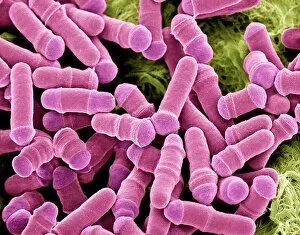
Pseudomonas aeruginosa bacteria, coloured scanning electron micrograph (SEM). These Gram-negative rod-shaped bacteria are found in soil and water, and as normal flora in the human intestine

Background imageFalse Coloured Collection: E. coli bacterium, TEM

E. coli bacterium, TEM
E. coli bacterium, coloured transmission electron micrograph (TEM). E. coli are Gram-negative rod-shaped bacteria that are part of the normal flora of the human gut

Background imageFalse Coloured Collection: Geranium pollen, SEM

Geranium pollen, SEM
Geranium pollen. Coloured scanning electron micrograph (SEM) of pollen (pink) on the stamen of a geranium flower (Geranium sp.)

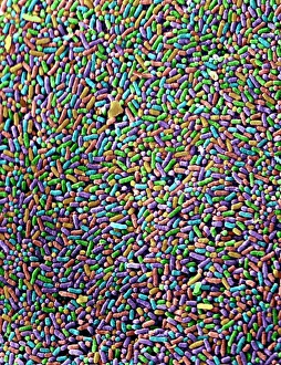
Background imageFalse Coloured Collection: Bacteria found on mobile phone, SEM

Bacteria found on mobile phone, SEM
Bacteria found on mobile phone. Coloured scanning electron micrograph (SEM) of bacteria cultured from a mobile phone. Tests have revealed the average handset carries 18 times more potentially harmful

Background imageFalse Coloured Collection: The four humours, 16th century artwork

The four humours, 16th century artwork
The four humours, coloured historical artwork. Here, the head of Christ is surrounded by four male figures, who are personifications of the four humours

Background imageFalse Coloured Collection: Gregor Mendel, Austrian botanist

Gregor Mendel, Austrian botanist
Gregor Johann Mendel (1822-1884), Austrian botanist and founder of genetics. Mendel, the abbot of an abbey in Brno, carried out breeding experiments with pea plants (held in hand)

Background imageFalse Coloured Collection: Blood clot, SEM

Blood clot, SEM
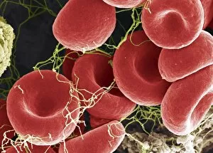
Blood clot, coloured scanning electron micrograph (SEM). Red blood cells are red and fibrin protein strands are green. Platelets are at bottom right

Background imageFalse Coloured Collection: Dermestid beetle, SEM

Dermestid beetle, SEM
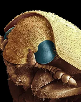
Dermestid beetle. Scanning electron micrograph (SEM(=) of a dermestid beetle. Dermestidae are a family of Coleoptera that are commonly referred to as skin beetles

Background imageFalse Coloured Collection: 19th-century gold mining, Australia

19th-century gold mining, Australia. Artwork of Chinese workers excavating and washing gold ores at a gold mine in Australia

Background imageFalse Coloured Collection: Medical zodiac, 15th century diagram

Medical zodiac, 15th century diagram
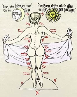
Medical zodiac. 15th century diagram with Latin text illustrating how the human body relates to the zodiac signs. Such information was used in medical practices, such as bloodletting

Background imageFalse Coloured Collection: Galileo and his daughter Maria Celeste

Galileo and his daughter Maria Celeste
Galilei Galileo (1564-1642), Italian physicist and astronomer, being guided by his daughter Maria Celeste (1600-1634), a nun

Background imageFalse Coloured Collection: Normal spine, X-ray

Normal spine, X-ray
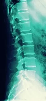
Normal spine. Coloured profile X-ray showing bones of the spine (vertebrae, centre). The front of the body is at right. The ribs (upper right) and the pelvis (bottom) are also seen

Background imageFalse Coloured Collection: Tendon, SEM

Tendon, SEM
Tendon, coloured scanning electron micrograph (SEM), showing bundles of collagen fibres. Magnification: x5000 when printed at 10 centimetres wide

Background imageFalse Coloured Collection: Macrophage engulfing TB bacteria, SEM

Macrophage engulfing TB bacteria, SEM
Macrophage engulfing TB bacteria. Coloured scanning electron micrograph (SEM) of a macrophage white blood cell (purple) engulfing a tuberculosis (Mycobacterium tuberculosis) bacterium (pink)

Background imageFalse Coloured Collection: Oriental stargazer lily (Lilium sp. ), coloured X-ray

Oriental stargazer lily (Lilium sp. ), coloured X-ray
Oriental stargazer lily (Lilium sp.), coloured X-ray

Background imageFalse Coloured Collection: Leg in stiletto shoe MRI style, X-ray

Leg in stiletto shoe MRI style, X-ray
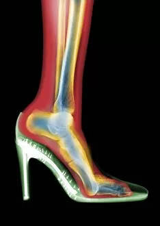
Leg in stiletto shoe, coloured MRI style X-ray

Background imageFalse Coloured Collection: Needle playing a record, SEM

Needle playing a record, SEM
Needle playing a record. Coloured scanning electron micrograph (SEM) of the needle (stylus) of a record player in a groove on a record. A record is used to store sound

Background imageFalse Coloured Collection: Ragworm mouth, SEM

Ragworm mouth, SEM
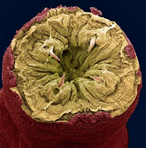
Ragworm mouth. Coloured scanning electron micrograph (SEM) of the mouth of a ragworm (Nereis sp.). Four teeth (cream) are seen around the opening. These worms are used commercially as fishing bait

Background imageFalse Coloured Collection: Black Death rat flea, artwork

Black Death rat flea, artwork
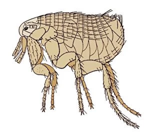
Oriental rat flea (Xenopsylla cheopis), artwork. This flea was the primary vector of the bacterium Yersinia pestis, which caused the Black Death

Background imageFalse Coloured Collection: Head of a young newt, SEM

Head of a young newt, SEM
Head of a young newt. Scanning electron micrograph (SEM) of a juvenile (immature) newt. Although this newt is in the last stages of development

Background imageFalse Coloured Collection: Kidney glomeruli, SEM

Kidney glomeruli, SEM
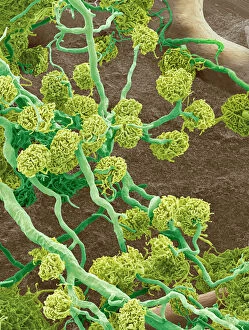
Kidney glomeruli, coloured scanning electron micrograph (SEM). The cells that usually surround the glomeruli have been removed to reveal the coiled capillaries at the core of the glomeruli

Background imageFalse Coloured Collection: Nerve cell, TEM

Nerve cell, TEM
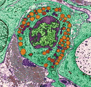
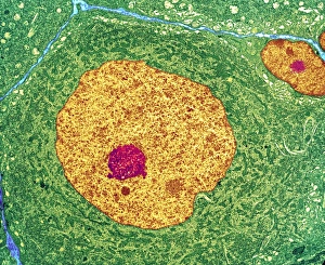
Nerve cell. Coloured transmission electron micrograph (TEM) of a nerve cell body in cross- section. The cell has a large nucleus (yellow) and inner nucleolus (red)

Background imageFalse Coloured Collection: Red blood cells, SEM

Red blood cells, SEM
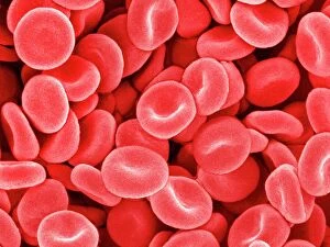
Red blood cells, coloured scanning electron micrograph (SEM). Red blood cells (erythrocytes) are biconcave, giving them a large surface area for gas exchange, and highly elastic

Background imageFalse Coloured Collection: Phagocytosis of fungal spores, SEM

Phagocytosis of fungal spores, SEM
Phagocytosis of a fungus spore. Coloured scanning electron micrograph (SEM) of a neutrophil (blue) phagocytosing (engulfing and destroying) spores from the fungus Aspergillus fumigatus (red)


All Professionally Made to Order for Quick Shipping

"Revealing the Unseen: Exploring the World Through False Colors" Step back in time to medieval alchemy, where a peculiar contraption known as the "Medieval urine wheel" was used to extract hidden secrets from bodily fluids. Fast forward to modern times and witness how science has evolved, unveiling breathtaking wonders like the Orion Nebula. Delve into the microscopic realm, where particle tracks paint a mesmerizing picture of subatomic interactions. Journey across Martian landscapes through the eyes of Spirit rover images, capturing otherworldly vistas that ignite our imagination. Peering into our own genetic blueprint, false colors bring clarity to X and Y chromosomes, unraveling mysteries within our very cells. A horse's skull takes on an ethereal glow as we explore its intricate structure using advanced imaging techniques. Witness synapse nerve junctions come alive with vibrant hues under a transmission electron microscope (TEM), revealing connections that shape our thoughts and actions. A person holding a camera becomes an enigma when captured through X-ray technology – their inner world exposed for all to see. Marvel at tardigrades' resilience as they navigate their microscopic universe under scanning electron microscopy (SEM). Calcareous phytoplankton dance with color in intricate patterns while nerve cells reveal their complexity in stunning detail. Travel back in time to 19th-century Cornwall and discover the harsh reality of tin mining brought vividly to life through false colors – reminding us of humanity's enduring spirit even amidst adversity. In this captivating journey through various realms – from ancient alchemy to distant galaxies, minuscule particles to majestic landscapes – false colors unlock hidden dimensions beyond what meets the eye, inviting us all into a world brimming with wonder and discovery.