
Fruiting Body Collection (page 2)
"Fruiting Body: A Fascinating World of Mushroom and Slime Moulds" Nature's artistic creations never cease to amaze us


All Professionally Made to Order for Quick Shipping
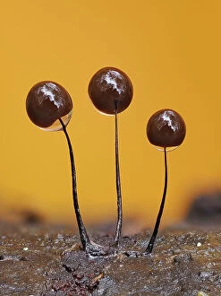
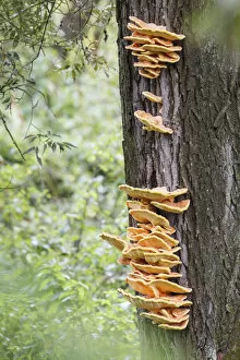
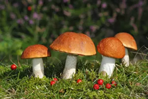
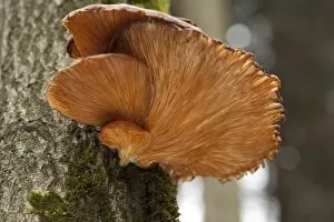
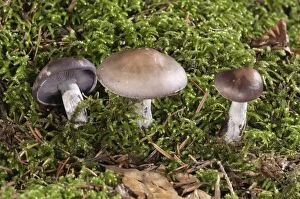
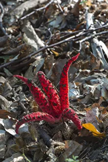
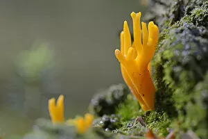
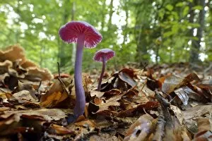
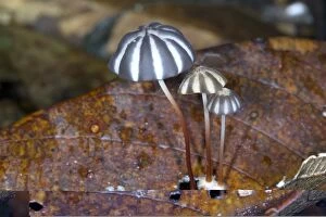
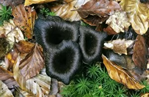
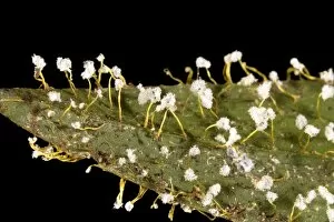
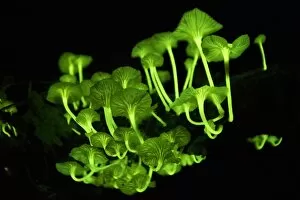
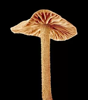
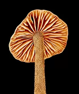
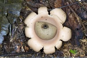
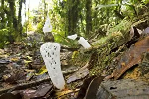
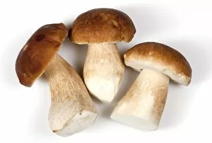
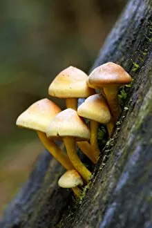
"Fruiting Body: A Fascinating World of Mushroom and Slime Moulds" Nature's artistic creations never cease to amaze us, especially when it comes to the diverse forms and structures found in fruiting bodies. From the iconic Fly agaric mushrooms with their vibrant red caps speckled with white dots, to the intricate patterns of mushroom gills observed under a scanning electron microscope (SEM), these organisms captivate our imagination. But it doesn't stop there. The world of fruiting bodies extends beyond mushrooms alone. Take, for instance, the fruiting bodies of Rhizopus oligosporus, a type of mold used in food fermentation processes like tempeh production. These tiny structures play a crucial role in transforming ingredients into delicious culinary delights. Moving on from molds to lichens, we encounter the enchanting Cup lichen (Cladonia floerkeana). Its cup-shaped fruiting body adds an ethereal touch to forest floors or rocky landscapes where it thrives. And then there are slime moulds – peculiar organisms that blur the line between fungi and protists. In Buckinghamshire, England's woodlands during November, Stemonitopsis typhina sporangia can be spotted growing on bark edges. These slimy masses release spores as they mature—a mesmerizing sight captured through focus stacking photography techniques. Another slime mould species found in Buckinghamshire is Metatrichia floriformis. Its line of split-open sporangia reveals delicate spore-filled interiors against a backdrop of winter scenery—nature's own miniature explosions frozen in time. Zooming even closer into this microscopic world brings us Lamproderma scintillans—a slime mould boasting 1mm tall sporangia that shimmer like stars under magnification—an awe-inspiring spectacle hidden within nature's nooks and crannies.