Fraud Blocker Skip to main content

Fruiting Body Collection (page 4)

"Fruiting Body: A Fascinating World of Mushroom and Slime Moulds" Nature's artistic creations never cease to amaze us

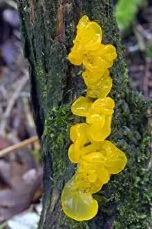
Background imageFruiting Body Collection: Tremella mesenterica

Tremella mesenterica
Yellow brain fungus (Tremella mesenterica) growing on a dead gorse bush. The fruiting body is soft, gelatinous and translucent with brain like lobes

Background imageFruiting Body Collection: Common Earthball (Scleroderma citrinum)

Common Earthball (Scleroderma citrinum). The fruiting body is hemispherical with a tough outer layer covered in scales. Inside the ball is a purple-brown mass (gleba)

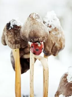
Background imageFruiting Body Collection: Ladybird on fungus

Ladybird on fungus. Seven-spot ladybird (Coccinella septem-punctata) climbing up the stalk of a fungal fruiting body. This is a wood-rotting fungus growing on a dead tree stump

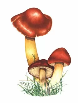
Background imageFruiting Body Collection: Crimson waxcap mushrooms, artwork

Crimson waxcap mushrooms, artwork
Crimson waxcap (Hygrocybe punicea) mushrooms, artwork. This fungus is found in northern Europe. The mushrooms (fruiting bodies) have a bell-shaped cap (lower right)

Background imageFruiting Body Collection: Parasol mushroom

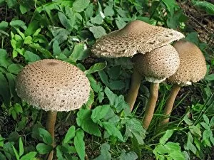
Parasol mushroom (Macrolepiota procera). Photographed on Powerstock Common, Dorset, UK, in October

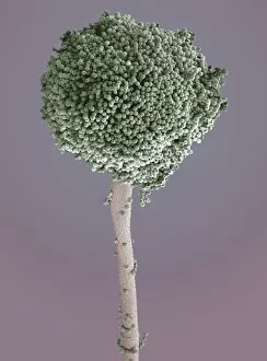
Background imageFruiting Body Collection: Aspergillus fungus, SEM

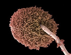
Aspergillus fungus, SEM
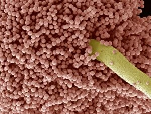
Aspergillus fungus. Coloured scanning electron micrograph (SEM) of the fruiting body (round, top) of a typical mould (Aspergillus sp.)

Background imageFruiting Body Collection: Woman holding split puffball fungus

Woman holding split puffball fungus
MODEL RELEASED. Woman holding a split puffball fungus. The outer wall of this fruiting body has split open to allow the dispersal of the reproductive spores from the sac inside

Background imageFruiting Body Collection: Curvularia geniculata fungus, SEM

Curvularia geniculata fungus, SEM
Curvularia geniculata fungus. Coloured scanning electron micrograph (SEM) of the fruiting body of the mould fungus Curvularia geniculata

Background imageFruiting Body Collection: Bracket fungus (Laetiporus sulphureus)

Bracket fungus (Laetiporus sulphureus)
Bracket fungus. Young sulphur-coloured fruiting body of the edible bracket fungus known as the chicken of the woods (Laetiporus sulphureus), growing on a living willow tree

Background imageFruiting Body Collection: Bracket fungus (Daedaleopsis confragosa)

Bracket fungus (Daedaleopsis confragosa)
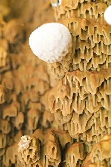
Blushing bracket fungus (Daedaleopsis confragosa). Close-up of the underside of the blushing bracket fungus showing its pores. The pores are the ends of the tubes within which the spores are produced

Background imageFruiting Body Collection: Bracket fungus (Polyporus squamosus)

Bracket fungus (Polyporus squamosus)
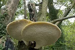
Bracket fungus. Dryads saddle (Polyporus squamosus) bracket fungus on a tree

Background imageFruiting Body Collection: Parasol mushrooms

Parasol mushrooms (Macrolepiota procera). Photographed in Narke, Sweden

Background imageFruiting Body Collection: Psathyrella mushrooms after forest fire

Psathyrella mushrooms after forest fire
Psathyrella pennata mushrooms growing one year after a forest fire. This fire took place in a clear-felled area in the Tiveden forest, in Vastra Gotaland County, Sweden

Background imageFruiting Body Collection: Geopyxis elf-cup fungi after forest fire

Geopyxis elf-cup fungi after forest fire
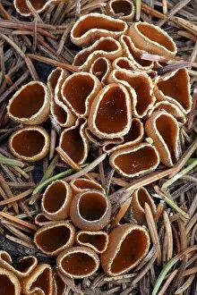
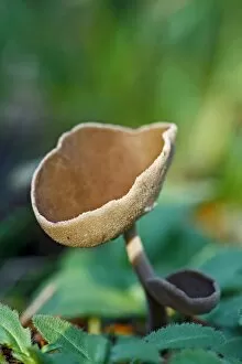
Geopyxis carbonaria elf-cup fungi growing one year after a forest fire. This fire took place in a clear-felled area in the Tiveden forest, in Vastra Gotaland County, Sweden

Background imageFruiting Body Collection: Reishi fungus

Reishi fungus
Reishe fungus (Ganoderma lucidum) growing on a dead tree. Photographed in Narke, Sweden

Background imageFruiting Body Collection: Honey fungus

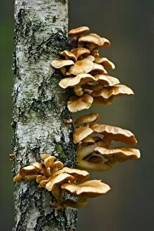
Honey fungus (Armillaria mellea) growing on a tree trunk. Photogroahed in Narke, Sweden

Background imageFruiting Body Collection: Jersey cow bolete

Jersey cow bolete (Suillus bovinus). Photographed in Narke, Sweden

Background imageFruiting Body Collection: Myxomphalia mushroom after forest fire

Myxomphalia mushroom after forest fire
Myxomphalia maura mushrooms growing one year after a forest fire. This fire took place in a clear-felled area in the Tiveden forest, in Vastra Gotaland County, Sweden

Background imageFruiting Body Collection: Helvella macropus mushroom

Helvella macropus mushroom
Helvella macropus fungus fruiting bodies. Photographed in Narke, Sweden
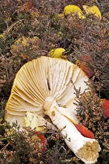
Background imageFruiting Body Collection: Hares ear fungus

Hares ear fungus (Otidea onotica). Photographed in Narke, Sweden

Background imageFruiting Body Collection: Peziza cup fungus after forest fire

Peziza cup fungus after forest fire
Peziza cup fungus (Peziza lobulata) growing two months after a forest fire. This fire took place in a clear-felled area in the Tiveden forest, in Vastra Gotaland County, Sweden

Background imageFruiting Body Collection: Plant regrowth after forest fire

Plant regrowth after forest fire
Plant regrowth one year after a forest fire. The plants and fungi seen here include Pholiota highlandensis mushrooms (lower left), the common liverwort (Marchantia polymorpha, white-green, centre)

Background imageFruiting Body Collection: Birch bolete (Leccinum scabrum) mushroom

Birch bolete (Leccinum scabrum) mushroom
Brown birch bolete (Leccinum scabrum) mushroom growing amongst common heather (Calluna vulgaris). Photographed in the Cairngorms National Park, Scotland, in August

Background imageFruiting Body Collection: Mushroom gill cap, light micrograph

Mushroom gill cap, light micrograph. Transverse section through the cap of a common mushroom (Psalliota sp.) showing the gill lamellae (white)

Background imageFruiting Body Collection: Fly agaric (Amanita muscaria) mushrooms

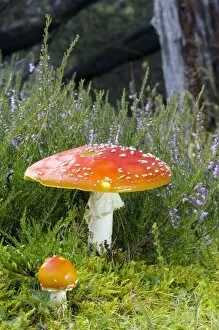
Fly agaric (Amanita muscaria) mushrooms growing amongst common heather (Calluna vulgaris) in an ancient Caledonian Scots Pine woodland

Background imageFruiting Body Collection: Mould fruiting bodies, light micrograph

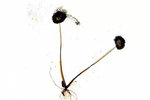
Mould fruiting bodies, light micrograph. Black mould (Rhizopus nigricans) fruiting bodies (top and upper right) liberating spores

Background imageFruiting Body Collection: Aspergillus fungus spores, SEM

Aspergillus fungus spores, SEM
Aspergillus fungus spores, coloured scanning electron micrograph (SEM). These chains of spores (orange) are the reproductive cells of the fruiting body, or conidiophore

Background imageFruiting Body Collection: Amanita (Amanita frostiana) mushroom

Amanita (Amanita frostiana) mushroom
Frosts amanita (Amanita frostiana) mushroom. Photographed in Vermont, USA

Background imageFruiting Body Collection: Woolly gomphus (Gomphus floccosus)

Woolly gomphus (Gomphus floccosus) mushroom. Photographed in Vermont, USA

Background imageFruiting Body Collection: Dry rot fungus

Dry rot fungus. Dry rot, unlike its name suggests, infects damp timber. The term refers to the growth of either of two fungi

Background imageFruiting Body Collection: Bleeding mycena mushrooms

Bleeding mycena mushrooms (Mycena haemotopus). Bleeding mycenas are so called because they release a reddish juice when cut. They grow on rotting wood, reaching about 3-8 centimetres in height

Background imageFruiting Body Collection: Earth star mushroom

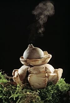
Earth star mushroom (Geastrum triplex) growing in woodland soil. It has released (puffed) a cloud of reproductive spores (upper centre)

Background imageFruiting Body Collection: Mushroom surface, SEM

Mushroom surface, SEM
Mushroom surface. Coloured scanning electron micrograph (SEM) of the spore-bearing surface of a hen of the woods mushroom (Grifola frondosa). This surface is found on the underside of the mushroom

Background imageFruiting Body Collection: Mushroom gills

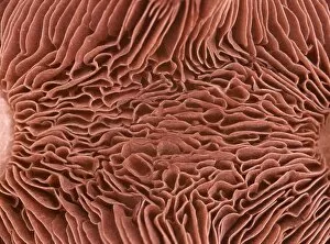
Mushroom gills. Macrophotograph of the gills of an unidentified double capped mushroom. Gills are found on the underside of the cap (fruiting body) and hold the fungal spores

Background imageFruiting Body Collection: Fly agaric (Amanita muscaria)

Fly agaric (Amanita muscaria) mushroom lying among ling heather (Calluna vulgaris). This fungus is poisonous if ingested, however it is often used for its hallucinogenic properties when used in small

Background imageFruiting Body Collection: Bog beacon fungus (Mitrula paludosa)

Bog beacon fungus (Mitrula paludosa)
Bog beacon fungus (Mitrula paludosa, orange) amongst sphagnum moss in a bog. Photographed in the Cairngorms National Park, Aberdeenshire, Scotland, in June

Background imageFruiting Body Collection: Oyster mushrooms

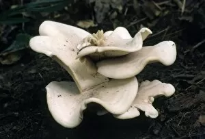
Oyster mushrooms (Pleurotus ostreatus) growing on a tree trunk. These fruiting bodies (reproductive structures) are produced year-round by the fungus in large clusters

Background imageFruiting Body Collection: Crimson wax cap mushroom

Crimson wax cap mushroom (Hygrocybe punicea). Photographed in the New Forest, UK

Background imageFruiting Body Collection: Stinky squid fungus

Stinky squid fungus

Background imageFruiting Body Collection: Pipe club fungus

Pipe club fungus (Clavariadelphus fistulosus) on a tree branch

Background imageFruiting Body Collection: Bracket fungus

Bracket fungus (class Basidiomycetes). This fungus forms these fruiting bodies to spread its reproductive spores through surface pores. Photographed in Vermont, USA

Background imageFruiting Body Collection: Honey fungus mushrooms

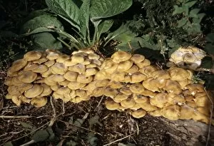
Honey fungus mushrooms or bootlace mushrooms (Armillaria mellea). This fungus is found on or around the trunks of coniferous and deciduous trees

Background imageFruiting Body Collection: Fungus (Lepista caespitose)

Fungus (Lepista caespitose). Photographed in Russia

Background imageFruiting Body Collection: Fungal spores, SEM

Fungal spores, SEM
Fungal spores. Coloured scanning electron micrograph (SEM) of fungal spores on a fruiting body of an Aspergillus sp. fungus. The name for such a fruiting body is a conidiophore
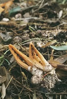
Background imageFruiting Body Collection: Fruiting bodies of fungus, Cordyceps

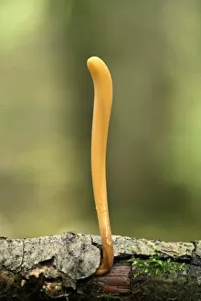
Fruiting bodies of fungus, Cordyceps
Parasitic fungus. Fruiting bodies from a parasitic fungus of the Cordyceps sp. Three brown club-like fruiting bodies are seen emerging from the forest ground

Background imageFruiting Body Collection: Parasol mushrooms, Lepiota procera

Parasol mushrooms, Lepiota procera
Parasol mushrooms. The fruiting bodies of Lepiota procera, the Parasol mushroom. This large mushroom produces a flattened pale brown cap of up to 25cm diameter

Background imageFruiting Body Collection: Fly Agaric funghi in Windermere Cumbria UK

Fly Agaric funghi in Windermere Cumbria UK
Ashley Cooper / SpecialistStock


All Professionally Made to Order for Quick Shipping

"Fruiting Body: A Fascinating World of Mushroom and Slime Moulds" Nature's artistic creations never cease to amaze us, especially when it comes to the diverse forms and structures found in fruiting bodies. From the iconic Fly agaric mushrooms with their vibrant red caps speckled with white dots, to the intricate patterns of mushroom gills observed under a scanning electron microscope (SEM), these organisms captivate our imagination. But it doesn't stop there. The world of fruiting bodies extends beyond mushrooms alone. Take, for instance, the fruiting bodies of Rhizopus oligosporus, a type of mold used in food fermentation processes like tempeh production. These tiny structures play a crucial role in transforming ingredients into delicious culinary delights. Moving on from molds to lichens, we encounter the enchanting Cup lichen (Cladonia floerkeana). Its cup-shaped fruiting body adds an ethereal touch to forest floors or rocky landscapes where it thrives. And then there are slime moulds – peculiar organisms that blur the line between fungi and protists. In Buckinghamshire, England's woodlands during November, Stemonitopsis typhina sporangia can be spotted growing on bark edges. These slimy masses release spores as they mature—a mesmerizing sight captured through focus stacking photography techniques. Another slime mould species found in Buckinghamshire is Metatrichia floriformis. Its line of split-open sporangia reveals delicate spore-filled interiors against a backdrop of winter scenery—nature's own miniature explosions frozen in time. Zooming even closer into this microscopic world brings us Lamproderma scintillans—a slime mould boasting 1mm tall sporangia that shimmer like stars under magnification—an awe-inspiring spectacle hidden within nature's nooks and crannies.