Fraud Blocker Skip to main content

Genes Collection (page 2)

"Unlocking the Secrets: Exploring the Fascinating World of Genes" The X and Y chromosomes: Unraveling the Blueprint of Life

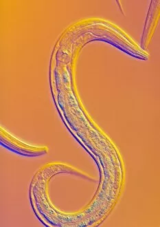
Background imageGenes Collection: C. elegans worm

C. elegans worm
Caenorhabditis elegans nematode worm, light micrograph. This soil-dwelling hermaphrodite nematode worm is one of the most studied animals in biological and genetic research

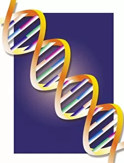
Background imageGenes Collection: DNA

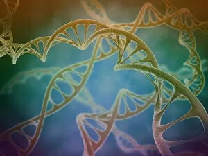
DNA, computer artwork. DNA (deoxyribonucleic acid) consists of two strands (yellow) of sugar phosphates forming a double helix

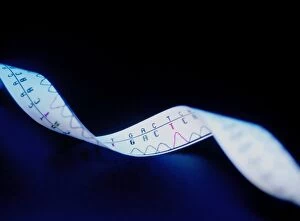
Background imageGenes Collection: Genetic sequence

Genetic sequence. Printout of the genetic code of a single strand of DNA (deoxyribonucleic acid). DNA normally comprises two spiralling paired strands of sugar phosphates that are linked by

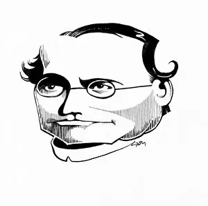
Background imageGenes Collection: Gregor Mendel, caricature

Gregor Mendel, caricature
Gregor Mendel (1822-1884). Caricature of the Austrian botanist and founder of genetics Gregor Johann Mendel. Mendel, the abbot of an abbey in Brno, Austria

Background imageGenes Collection: Uniforms civil guard Courtray Belgium 1832 Black chalk

Uniforms civil guard Courtray Belgium 1832 Black chalk
Artokoloro

Background imageGenes Collection: Grand Review Juste-Milieu Passing La Caricature

Grand Review Juste-Milieu Passing La Caricature
Artokoloro

Background imageGenes Collection: Prise de Constantine 19th century Pen ink watercolor

Prise de Constantine 19th century Pen ink watercolor
Artokoloro

Background imageGenes Collection: Album Lithographique 1830-1837 1830-1837 Overall

Album Lithographique 1830-1837 1830-1837 Overall
Artokoloro

Background imageGenes Collection: People Delivered Vampire Taxes 1833 Lithograph

People Delivered Vampire Taxes 1833 Lithograph
Artokoloro

Background imageGenes Collection: Conceptual image of a telomere

Conceptual image of a telomere. A telomere is a region of the DNA sequence at the end of a chromosome. Their function is to protect the ends of the chromosome from degradating

Background imageGenes Collection: Cell nucleus with chromosome

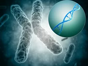
Cell nucleus with chromosome. The cell nucleus helps control eating, movement, and reproduction

Background imageGenes Collection: Microscopic view of DNA binding

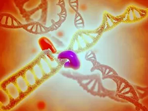
Microscopic view of DNA binding

Background imageGenes Collection: Microscopic view of telomeres highlighted at the tips of chromosome

Microscopic view of telomeres highlighted at the tips of chromosome. A telomere is a region of the DNA sequence at the end of a chromosome

Background imageGenes Collection: Microscopic view of DNA

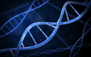
Microscopic view of DNA

Background imageGenes Collection: Conceptual image of chromosomes inside the blood stream

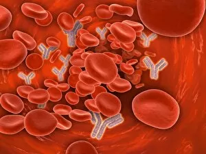
Conceptual image of chromosomes inside the blood stream

Background imageGenes Collection: Conceptual image of a telomere showing DNA structure

Conceptual image of a telomere showing DNA structure. A telomere is a region of the DNA sequence at the end of a chromosome. Their function is to protect the ends of the chromosome from degradation

Background imageGenes Collection: Microscopic view of chromosome

Microscopic view of chromosome

Background imageGenes Collection: Stylized view of strands of human DNA

Stylized view of strands of human DNA or deoxyribonucleic acid

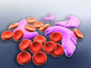
Background imageGenes Collection: Conceptual image of sickle cell anemia

Conceptual image of sickle cell anemia. Sickle cell anemia is a disease in which your body produces abnormally shaped red blood cells. The cells are shaped like a crescent or sickle

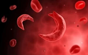
Background imageGenes Collection: Microscopic view of sicke cells causing anemia disease

Microscopic view of sicke cells causing anemia disease

Background imageGenes Collection: Microscopic view of cancer cells

Microscopic view of cancer cells. Cancer occurs when a cells gene mutations make the cell unable to correct DNA damage

Background imageGenes Collection: Conceptual image of chromosome

Conceptual image of chromosome

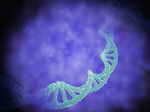
Background imageGenes Collection: Conceptual image of DNA

Conceptual image of DNA

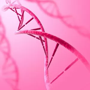
Background imageGenes Collection: Cluster of DNA strands

Cluster of DNA strands of human DNA or deoxyribonucleic acid

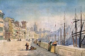
Background imageGenes Collection: The Port of Genes (Genoa), 1878. Watercolour by J. L Genatto. Sunlit buildings at quayside

The Port of Genes (Genoa), 1878. Watercolour by J. L Genatto. Sunlit buildings at quayside
The Port of Genes (Genoa), 1878. Watercolour by J.L Genatto. Sunlit buildings at quayside, women leaning on sea wall by masts of vessels docked vessels

Background imageGenes Collection: Sheep farming, shepherd using sterile single use pin on Texel ram nose to extract blood for

Sheep farming, shepherd using sterile single use pin on Texel ram nose to extract blood for Scrapie genotype testing, England, May

Background imageGenes Collection: Double Helix of Human DNA

Double Helix of Human DNA

Background imageGenes Collection: Artwork of DNA structure

Artwork of DNA structure

Background imageGenes Collection: Lion-jaguar hybrid cub (Panthera hybrid), side view

Lion-jaguar hybrid cub (Panthera hybrid), side view

Background imageGenes Collection: Stochastic gene expression, illustration C018 / 0906

Stochastic gene expression, illustration C018 / 0906
Stochastic gene expression, illustration. Every cell in an organism contains every single gene that makes up the organisms genome. However, they are not all active (expressed) in each cell
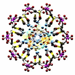
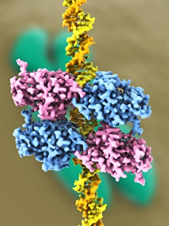
Background imageGenes Collection: Tumour suppressor protein and DNA C017 / 3647

Tumour suppressor protein and DNA C017 / 3647
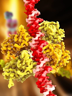
Tumour suppressor protein and DNA. Computer artwork showing a molecule of the tumour suppressor protein p53 (blue and pink) bound to a molecule of DNA (deoxyribonucleic acid, yellow and orange)

Background imageGenes Collection: DNA molecule, artwork C017 / 7217

DNA molecule, artwork C017 / 7217
DNA molecule. Computer artwork showing a double stranded DNA (deoxyribonucleic acid) molecule. DNA is composed of two strands twisted into a double helix

Background imageGenes Collection: DNA molecule, artwork C017 / 0616

DNA molecule, artwork C017 / 0616
DNA molecule. Computer artwork looking along the interior of a double stranded DNA (deoxyribonucleic acid) molecule. DNA is composed of two strands twisted into a double helix

Background imageGenes Collection: DNA molecule, artwork C017 / 0615

DNA molecule, artwork C017 / 0615
DNA molecule. Computer artwork looking along the interior of a double stranded DNA (deoxyribonucleic acid) molecule. DNA is composed of two strands twisted into a double helix

Background imageGenes Collection: DNA molecule, artwork C017 / 0617

DNA molecule, artwork C017 / 0617
DNA molecule. Computer artwork looking along the interior of a double stranded DNA (deoxyribonucleic acid) molecule. DNA is composed of two strands twisted into a double helix

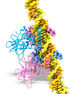
Background imageGenes Collection: Tumour suppressor protein and DNA C017 / 3644

Tumour suppressor protein and DNA C017 / 3644
Tumour suppressor protein and DNA. Computer artwork showing a molecule of the tumour suppressor protein p53 (blue and pink) bound to a molecule of DNA (deoxyribonucleic acid, yellow and orange)

Background imageGenes Collection: Tumour suppressor protein and DNA C017 / 3646

Tumour suppressor protein and DNA C017 / 3646
Tumour suppressor protein and DNA. Computer artwork showing a molecule of the tumour suppressor protein p53 (blue and pink) bound to a molecule of DNA (deoxyribonucleic acid, yellow and orange)

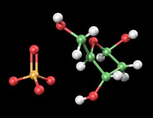
Background imageGenes Collection: DNA components, artwork C017 / 7350

DNA components, artwork C017 / 7350
DNA components. Computer artwork showing the structure of the two molecules that make up the backbone of DNA (deoxyribonucleic acid), phosphate (left) and deoxyribose (right)

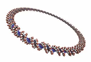
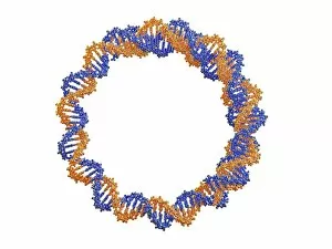
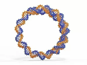
Background imageGenes Collection: Circular DNA molecule, artwork F006 / 7088

Circular DNA molecule, artwork F006 / 7088
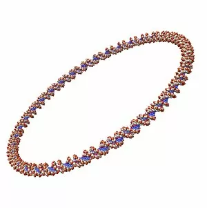
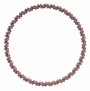
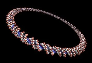
Circular DNA (deoxyribonucleic acid) molecule, computer artwork. Circular DNA has no ends, but consists of a ring structure

Background imageGenes Collection: Circular DNA molecule, artwork F006 / 7072

Circular DNA molecule, artwork F006 / 7072
Circular DNA (deoxyribonucleic acid) molecule, computer artwork. Circular DNA has no ends, but consists of a ring structure

Background imageGenes Collection: Circular DNA molecule, artwork F006 / 7095

Circular DNA molecule, artwork F006 / 7095
Circular DNA (deoxyribonucleic acid) molecule, computer artwork. Circular DNA has no ends, but consists of a ring structure

Background imageGenes Collection: Circular DNA molecule, artwork F006 / 7086

Circular DNA molecule, artwork F006 / 7086
Circular DNA (deoxyribonucleic acid) molecule, computer artwork. Circular DNA has no ends, but consists of a ring structure

Background imageGenes Collection: Circular DNA molecule, artwork F006 / 7083

Circular DNA molecule, artwork F006 / 7083
Circular DNA (deoxyribonucleic acid) molecule, computer artwork. Circular DNA has no ends, but consists of a ring structure

Background imageGenes Collection: Circular DNA molecule, artwork F006 / 7084

Circular DNA molecule, artwork F006 / 7084
Circular DNA (deoxyribonucleic acid) molecule, computer artwork. Circular DNA has no ends, but consists of a ring structure

Background imageGenes Collection: Glycine riboswitch molecule F007 / 9921

Glycine riboswitch molecule F007 / 9921
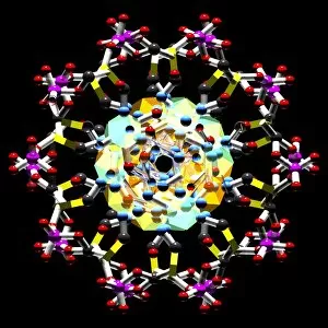
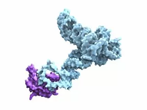
Molecular model of the bacterial glycine riboswitch. This is an RNA element that can bind the amino acid glycine. Glycine riboswitches usually consist of two metabolite-binding aptamer domains tandem

Background imageGenes Collection: Glycine riboswitch molecule F007 / 9906

Glycine riboswitch molecule F007 / 9906
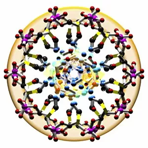
Molecular model of the bacterial glycine riboswitch. This is an RNA element that can bind the amino acid glycine. Glycine riboswitches usually consist of two metabolite-binding aptamer domains tandem

Background imageGenes Collection: Human chromosome

Human chromosome. Coloured scanning electron micrograph (SEM) of a human chromosome. Chromosomes occur in the nucleus of every cell in the body

Background imageGenes Collection: DNA molecule, artwork F008 / 2034

DNA molecule, artwork F008 / 2034
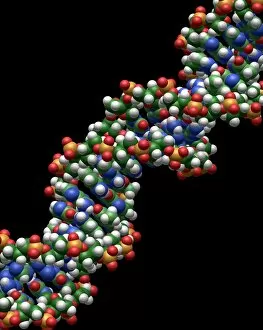
DNA molecule, computer artwork


All Professionally Made to Order for Quick Shipping

"Unlocking the Secrets: Exploring the Fascinating World of Genes" The X and Y chromosomes: Unraveling the Blueprint of Life. A mesmerizing sight: Leopard's black panther showcases melanistic phase, a result of its genes. DNA molecule: Nature's intricate code for life captured in stunning computer models. Abstract artwork reveals the beauty hidden within the DNA molecule. Gregor Mendel - Pioneering Austrian botanist who laid the foundation for understanding genetic inheritance. Peering into our origins: DNA Double Helix with Autoradiograph offers a glimpse into our genetic makeup. Guinea pigs showcase Mendel's Law through a vibrant poster, highlighting genetic patterns in action. Z-DNA tetramer molecule C015/6557 - Unveiling unique structures within our chromosomes. Chromosomes - The carriers of hereditary information that shape who we are. Delving deep into genetics, this captivating journey takes us from unraveling the mysteries held by X and Y chromosomes to witnessing nature's marvels like a leopard donning its striking black panther coat due to specific genes at play. The awe-inspiring complexity of life is encapsulated in DNA molecules, whether portrayed as computer models or abstract artworks that depict their elegance and intricacy. We pay homage to Gregor Mendel, an Austrian botanist whose groundbreaking work paved the way for understanding how traits are passed down through generations. Through images like DNA Double Helix with Autoradiograph or posters demonstrating Mendel's Laws using guinea pigs as examples, we gain insight into our own origins and witness firsthand how genetics shape every living being on this planet. Zooming further into microscopic wonders, we encounter Z-DNA tetramer molecules and explore fascinating structures found within our very own chromosomes – repositories of invaluable hereditary information.