Fraud Blocker Skip to main content

Glia Collection

Glia: The Unsung Heroes of the Nervous System Delicate and intricate

Background imageGlia Collection: Cerebellum tissue, light micrograph

Cerebellum tissue, light micrograph
Cerebellum tissue. Confocal light micrograph of a section through the cerebellum of the brain. Purkinje cells, a type of neuron (nerve cell), are red

Background imageGlia Collection: Hippocampus brain tissue

Hippocampus brain tissue
Hippocampus tissue. Light micrograph of a sagittal (side view) section through the hippocampus of the brain showing the nerve cells within it

Background imageGlia Collection: Glial stem cell culture, light micrograph

Glial stem cell culture, light micrograph
Glial stem cell culture. Fluorescent light micrograph of glial stem cells producing the protein NG2 (red) as they mature. These stem cells can differentiate into several types of glial cells

Background imageGlia Collection: Cerebellum tissue, light micrograph

Cerebellum tissue, light micrograph
Cerebellum tissue. Confocal light micrograph of a section through the cerebellum of the brain showing two types of glial cells (support cells); astrocytes (star-shaped)

Background imageGlia Collection: Cerebellum tissue, light micrograph

Cerebellum tissue, light micrograph
Cerebellum tissue. Confocal light micrograph of a section through the cerebellum of the brain. Purkinje cells, a type of neuron (nerve cell), are red

Background imageGlia Collection: Illustration of Oligodendrocyte glia cell or neuroglia

Illustration of Oligodendrocyte glia cell or neuroglia

Background imageGlia Collection: Illustration of an Astrocyte human glial cell

Illustration of an Astrocyte human glial cell

Background imageGlia Collection: Neurofibromatosis, a genetic disorder of the nervous system

Neurofibromatosis, a genetic disorder of the nervous system
A genetic disorder of the nervous system, neurofibromatosis causes tumors to form on nerves throughout the body, including a type of tumor called an optic nerve glioma that can result in childhood

Background imageGlia Collection: Directed differentiation of multipotential human neural progenitor cells

Directed differentiation of multipotential human neural progenitor cells
Human neural progenitor cells were isolated under selective culture conditions from the developing human brain and directed through lineage differentiation to GFAP + (glial fibrillary acid protein)

Background imageGlia Collection: Nervous system cells, artwork C017 / 3423

Nervous system cells, artwork C017 / 3423
Nervous system cells. Computer artwork showing neurons (nerve cells, purple), astrocytes (green) and a blood vessel (along right)

Background imageGlia Collection: Brain cell, TEM C014 / 0358

Brain cell, TEM C014 / 0358
Brain cell. Transmission electron micrograph (TEM) of a section through an oligodendrocyte in human brain tissue. Oligodendrocytes occur in both the white

Background imageGlia Collection: Brain cell, TEM C014 / 0359

Brain cell, TEM C014 / 0359
Brain cell. Transmission electron micrograph (TEM) of a section through an oligodendrocyte in human brain tissue. Oligodendrocytes occur in both the white

Background imageGlia Collection: Brain cells, TEM C013 / 4801

Brain cells, TEM C013 / 4801
Brain cells. Transmission electron micrograph (TEM) of a section through oligodendrocytes (dark) in human brain tissue, showing free ribosomes (dark green dots), golgi apparatus (curved brown lines)

Background imageGlia Collection: Brain cells, TEM C013 / 4800

Brain cells, TEM C013 / 4800
Brain cells. Transmission electron micrograph (TEM) of a section through oligodendrocytes in human brain tissue, showing free ribosomes (dark pink dots), golgi apparatus (curved brown lines)

Background imageGlia Collection: Brain cell, TEM C013 / 4799

Brain cell, TEM C013 / 4799
Brain cell. Transmission electron micrograph (TEM) of a section through an oligodendrocyte in human brain tissue, showing free ribosomes (dark brown dots), golgi apparatus (curved orange lines)

Background imageGlia Collection: Brain cell, TEM C013 / 4798

Brain cell, TEM C013 / 4798
Brain cell. Transmission electron micrograph (TEM) of a section through an oligodendrocyte in human brain tissue, showing free ribosomes (dark blue dots), golgi apparatus (curved light blue lines)

Background imageGlia Collection: Brain cortex tissue, light micrograph

Brain cortex tissue, light micrograph
Brain cortex tissue. Light micrograph of a section through tissue from the cortex of the brain. The cortex, or grey matter, is the outer layer of the brain

Background imageGlia Collection: Retina blood vessel and nerve cells

Retina blood vessel and nerve cells
Retina cells. Fluorescent light micrograph of cells in the retina, the light-sensitive membrane that lines the back of the eyeball

Background imageGlia Collection: Glial cells

Glial cells. Fluorescent light micrograph of glial cells from a mouse brain. The tissue has been tagged with fluorescent markers specific to certain proteins

Background imageGlia Collection: Human spinal cord, light micrograph

Human spinal cord, light micrograph

Background imageGlia Collection: Glial stem cell culture, light micrograph

Glial stem cell culture, light micrograph
Glial stem cell culture. Fluorescent light micrograph of glial stem cells producing the proteins NG2 (green) and OLIG2 (oligodendrocyte lineage transcription factor 2, red) as they mature

Background imageGlia Collection: Brain cells in culture, light micrograph

Brain cells in culture, light micrograph
Brain cells in culture. Fluorescent light micrograph of a microglial cell (upper left) and an oligodendrocyte (centre) from a human brain


All Professionally Made to Order for Quick Shipping

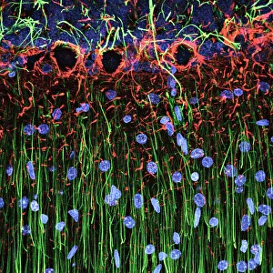
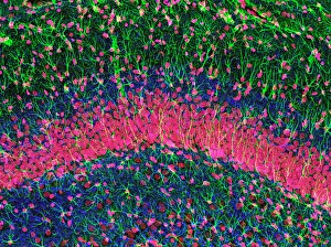
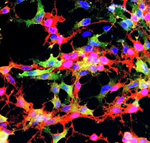
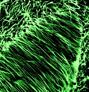
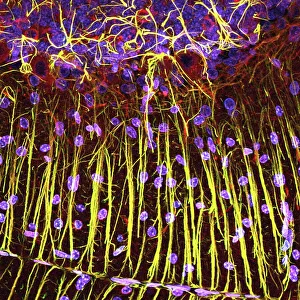
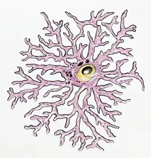
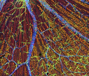
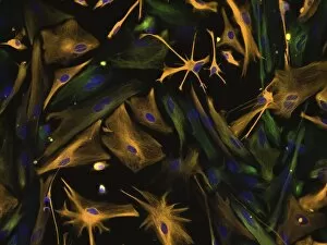
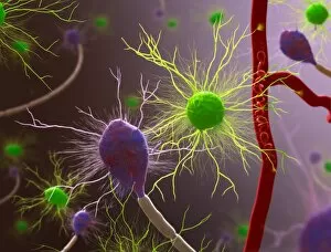
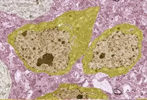
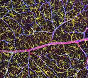
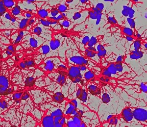
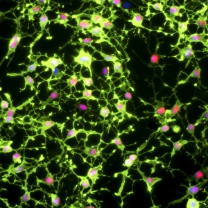
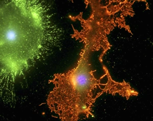
Glia: The Unsung Heroes of the Nervous System Delicate and intricate, the cerebellum tissue reveals a mesmerizing network of glial cells when observed under a light micrograph. In the hippocampus brain tissue, glia take center stage, supporting and nourishing neurons to ensure optimal brain function. A closer look at a light micrograph of glial stem cell culture unveils their remarkable ability to proliferate and differentiate into various specialized cells within the nervous system. Once again, the cerebellum tissue showcases its complexity as numerous glial cells are captured in another captivating light micrograph. Glia - often overshadowed by neurons - play an essential role in maintaining neuronal health and functionality, as depicted in this striking image of different types of glial cells. An illustration vividly portrays an oligodendrocyte glia cell or neuroglia responsible for forming myelin sheaths around nerve fibers, enabling efficient electrical signal transmission throughout the nervous system. Meet astrocytes – human glial cells that provide structural support to neurons while also regulating chemical balance within the brain; their importance cannot be overstated. Neurofibromatosis, a genetic disorder affecting the nervous system's development and function, highlights how crucial it is for our bodies to have properly functioning glial cells. Witnessing directed differentiation of multipotential human neural progenitor cells gives us hope for future treatments targeting specific neurological conditions with precision and efficacy. Through stunning artwork depicting various nervous system cells' intricacies (C017 / 3423), we gain insight into just how diverse and vital these cellular components truly are. Brain cell TEM C014 / 0358 offers a glimpse into one small part of our complex neural network where countless dedicated glial cells tirelessly work behind-the-scenes to maintain equilibrium. Glia - the unsung heroes of the nervous system.