Fraud Blocker Skip to main content

Helices Collection (page 7)

"Unraveling the Mysteries of Helices: Exploring the Intricate World of Molecular Structures" DNA Transcription Unveiled

Background imageHelices Collection: Bacteriorhodopsin, artwork

Bacteriorhodopsin, artwork
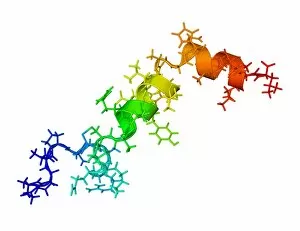
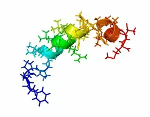
Bacteriorhodopsin. Artwork of the molecular structure of bacteriorhodopsin, a protein found in primitive micro-organisms known as Archaea

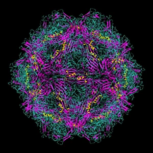
Background imageHelices Collection: Feline distemper virus particle

Feline distemper virus particle. Molecular model showing the structure of the capsid (outer protein coat) of a feline panleukopenia virus particle. This parovirus causes distemper in cats

Background imageHelices Collection: Cholesterol producing enzyme and statin

Cholesterol producing enzyme and statin. Computer model showing the molecular structure of HMG-CoA reductase (HMGCR) in complex with Atorvastatin

Background imageHelices Collection: Interferon gamma molecule and receptor

Interferon gamma molecule and receptor
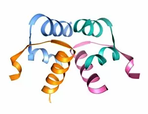
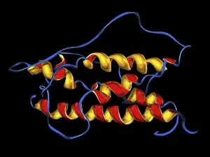
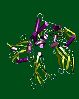
Interferon gamma. Molecular model showing the secondary structure of the interferon gamma dimer (bright pink and gold and pale pink and beige)

Background imageHelices Collection: Tobacco mosaic virus, molecular model

Tobacco mosaic virus, molecular model
Tobacco mosaic virus. Computer model showing the molecular structure of a tobacco mosaic virus (TMV) particle (virion). This plant virus is a tobamovirus, and has an RNA (ribonucleic acid) genome

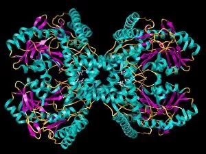
Background imageHelices Collection: Selenocysteine synthase enzyme molecule

Selenocysteine synthase enzyme molecule. Computer model showing the molecular structure of the enzyme selenocysteine synthase (SecS)

Background imageHelices Collection: Genetic biodiversity, conceptual image

Genetic biodiversity, conceptual image. Array of multi-coloured helices of the genetic molecule DNA (deoxyribonucleic acid)

Background imageHelices Collection: DNA replication, artwork

DNA replication, artwork
DNA replication. Computer artwork of a DNA (deoxyribonucleic acid) molecule replicating. DNA is composed of two strands twisted into a double helix

Background imageHelices Collection: Lysyl oxidase enzyme molecule

Lysyl oxidase enzyme molecule. Computer artwork showing the secondary structure of the enzyme lysyl oxidase (LOX). LOX is a homodimeric (composed of two identical subunits)

Background imageHelices Collection: Spanish flu H1 antigen, molecular model

Spanish flu H1 antigen, molecular model
Spanish flu virus antigen. Computer model showing the molecular structure of a haemagglutinin 1 (H1) antigen from the Spanish influenza (flu) virus particle

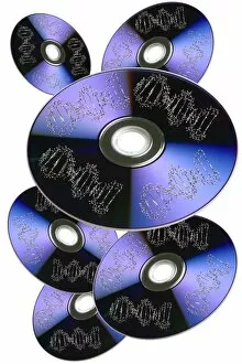
Background imageHelices Collection: Genetic information storage, artwork

Genetic information storage, artwork
Genetic information storage, conceptual computer artwork. Molecules of DNA (deoxyribonucleic acid) on the surface of dvds

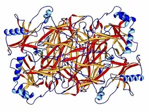
Background imageHelices Collection: Proteinase inhibitor molecule

Proteinase inhibitor molecule
Proteinase inhibitor. Molecular model showing the secondary structure of alpha-1 proteinase inhibitor, also known as alpha 1-antitrypsin

Background imageHelices Collection: Leptin hormone molecule

Leptin hormone molecule. Computer model showing the secondary structure of the hormone leptin. This hormone is produced by adipose (fat) tissue

Background imageHelices Collection: Inhibin beta A molecule

Inhibin beta A molecule. Computer model showing the crystal structure of a molecule of the protein Inhibin beta A (INHBA)

Background imageHelices Collection: Prolactin hormone molecule

Prolactin hormone molecule. Computer model showing the secondary structure of human prolactin (hPRL), or luteotropic hormone (LTH)

Background imageHelices Collection: Thrombopoietin hormone molecule

Thrombopoietin hormone molecule. Computer model showing the secondary structure of a molecule of the hormone thrombopoietin (TPO)

Background imageHelices Collection: Follicle stimulating hormone molecules

Follicle stimulating hormone molecules. Computer artwork showing the secondary structure of two strands of follicle stimulating hormone (FSH)

Background imageHelices Collection: Orexin-A hormone molecule

Orexin-A hormone molecule. Computer model showing the crystal structure of a molecule of the hormone orexin-A. The crystal structure consists of both the secondary structure

Background imageHelices Collection: Orexin-B hormone molecule

Orexin-B hormone molecule. Computer model showing the crystal structure of a molecule of the hormone orexin-B. The crystal structure consists of both the secondary structure

Background imageHelices Collection: Relaxin hormone molecule

Relaxin hormone molecule. Computer model showing the secondary structure of a molecule of the hormone relaxin. The alpha helices (ribbons) of the secondary structure can be seen

Background imageHelices Collection: Somatotrophin hormone molecule

Somatotrophin hormone molecule. Computer model showing the secondary structure of the human growth hormone somatotrophin (GH)

Background imageHelices Collection: Kinase molecule, secondary structure

Kinase molecule, secondary structure
Kinase molecule, computer model. Kinases are enzymes that catalyse the transfer of phosphate groups from a high-energy phosphate-containing molecule (such as ATP or ADP)

Background imageHelices Collection: Caspase 3 molecule

Caspase 3 molecule
Caspase-3 molecule. Computer artwork showing the secondary structure of a molecule of caspase-3. Caspase-3 is a protease, an enzyme that cleaves proteins

Background imageHelices Collection: Caspase 1 molecule

Caspase 1 molecule
Caspase-1 molecule. Computer artwork showing the secondary structure of a molecule of caspase-1. Caspase-1 is a protease, an enzyme that cleaves proteins

Background imageHelices Collection: Lipase molecule, secondary structure

Lipase molecule, secondary structure
Lipase molecule, computer model. Lipase is an enzyme that breaks lipids (fats) into fatty acids and glycerol. Human pancreatic lipase is the main enzyme responsible for breaking down fat in the human

Background imageHelices Collection: Cholinesterase enzyme

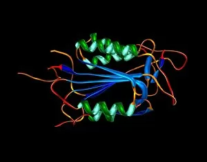
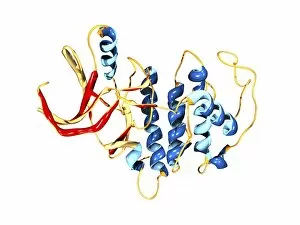
Cholinesterase enzyme. Molecular model of the secondary structure of butyrylcholinesterase (BChE), showing alpha helices (blue) and beta sheets (red and yellow)

Background imageHelices Collection: Ferroxidase enzyme, molecular model

Ferroxidase enzyme, molecular model
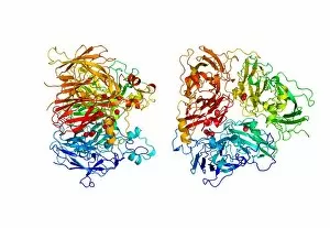
Ferroxidase enzyme. Molecular model showing two views of the secondary structure of the human enzyme ferroxidase, also known as ceruloplasmin. Copper atoms are represented as red spheres

Background imageHelices Collection: Synthetic peptide fibre, molecular model

Synthetic peptide fibre, molecular model
Synthetic peptide fibre. Molecular model of a synthetic collagen-like peptide fibre, showing three different ways of representing the structure. Peptides are small molecules formed from amino acids

Background imageHelices Collection: Viral dUTPase enzyme, molecular model

Viral dUTPase enzyme, molecular model
dUTPase enzyme. Molecular model of the enzyme dUTP pyrophosphatase (dUTPase) from the feline immunodeficiency virus (FIV)

Background imageHelices Collection: Cyclin-depenent kinase 5 molecule

Cyclin-depenent kinase 5 molecule
Cyclin-dependent kinase 5. Molecular model showing the secondary structure of the enzyme cylcin- dependent kinase 5 (cdk5)

Background imageHelices Collection: Protein from outer coat of SARS virus

Protein from outer coat of SARS virus
SARS virus capsid protein, molecular model. This protein is responsible for binding the capsid (outer coat) of the SARS (severe acute respiratory syndrome)

Background imageHelices Collection: Influenza A virus haemagglutinin protein

Influenza A virus haemagglutinin protein
Haemagglutinin protein from the surface of Influenza A virus, molecular model. This protein, known as haemagglutinin, is found on the surface of the influenza A virus

Background imageHelices Collection: Anthrax lethal factor protein

Anthrax lethal factor protein
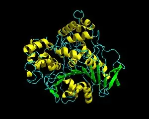
Anthrax lethal factor, molecular model. Lethal factor (LF) is one of the toxins produced by spores of the bacterium Bacillis anthracis

Background imageHelices Collection: Human growth hormone, molecular model

Human growth hormone, molecular model
Human growth hormone. Molecular model showing the secondary structure of human growth hormone (hGH), a hormone produced in the anterior pituitary gland in the brain

Background imageHelices Collection: Bacteriochlorophyll-containing protein

Bacteriochlorophyll-containing protein. Molecular models showing two views of the bacteriochlorophyll-containing protein found in the bacterium Prosthecochloris aestuarii

Background imageHelices Collection: FMD virus surface protein

FMD virus surface protein
Foot-and-mouth disease virus surface protein, molecular model. Foot-and-mouth disease (FMD) affects cloven-hooved mammals, commonly cattle and pigs

Background imageHelices Collection: Human RSV fusion core protein

Human RSV fusion core protein
Fusion core protein from the outer coat of the human respiratory syncytial virus (RSV), molecular model. RSV causes respiratory tract infections, usually with mild symptoms

Background imageHelices Collection: Antithrombin molecule

Antithrombin molecule
Antithrombin, molecular model. Antithrombin is a glycoprotein produced in the liver that inhibits enzymes involved in blood clotting

Background imageHelices Collection: Microglobulin protein, molecular model

Microglobulin protein, molecular model
Microglobulin protein. Molecular models of two overlapping views of the protein beta-2 microglobulin. This protein, with a relatively small molecular mass

Background imageHelices Collection: Bluetongue virus protein VP7 structure

Bluetongue virus protein VP7 structure
Bluetongue virus protein VP7, molecular model. VP7 is a protein forming part of the coating of the bluetongue virus. This virus, transmitted by midges

Background imageHelices Collection: Birch pollen allergen

Birch pollen allergen. Molecular model of the secondary structure of Bet v 1l, the molecule responsible for allergic reactions to birch pollen

Background imageHelices Collection: Cytochrome P450 molecule

Cytochrome P450 molecule
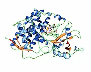
Cytochrome P450 protein. Molecular model showing the secondary structure of cytochrome P450. This protein plays a crucial role in metabolism in animals (including humans), fungi, plants and bacteria

Background imageHelices Collection: Reverse transcriptase enzyme from HIV

Reverse transcriptase enzyme from HIV, molecular model. This enzyme is from the human immunodeficiency virus (HIV) that causes AIDS

Background imageHelices Collection: Interleukin-10, molecular model

Interleukin-10, molecular model
Interleukin-10. Molecular model of the secondary structure of interleukin-10, a small protein known as a cytokine that plays an important regulatory role in the bodys immune system

Background imageHelices Collection: Mouse urinary protein, molecular model

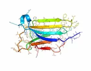
Mouse urinary protein, molecular model
Mouse urinary protein (MUP), molecular model. MUP refers to a family of similar proteins found in mouse urine. The proteins act as pheromones, airborne chemical signals

Background imageHelices Collection: Bluetongue virus protein structure

Bluetongue virus protein structure. Molecular model of some of the protein structures found in the protein coating of the bluetongue virus (BTV)

Background imageHelices Collection: Thrombin protein

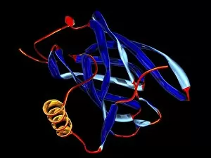
Thrombin protein

Background imageHelices Collection: Erythropoietin hormone

Erythropoietin hormone
Erythropoietin. Computer model of a molecule of the hormone erythropoietin. This is a glycoprotein that consists of a single-chain polypeptide of 165 amino acids


All Professionally Made to Order for Quick Shipping

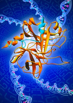
"Unraveling the Mysteries of Helices: Exploring the Intricate World of Molecular Structures" DNA Transcription Unveiled: Witness the intricate dance as DNA unravels and transcribes its genetic code, paving the way for life's blueprint. A Symphony in Proteins: Secondary structures adorned with helical motifs create a mesmerizing molecular model, showcasing nature's artistic prowess. Nucleosome Molecule - Nature's Genetic Sculptor: Behold the elegant architecture of nucleosomes, where DNA elegantly wraps around histone proteins forming a helical masterpiece. The Dance of Life: An artwork depicting a vibrant DNA molecule reveals its double-helix structure, symbolizing our very essence and genetic heritage. Bacterial Ribosome - The Protein Factory: Dive into the intricacies of bacterial ribosomes as they diligently translate genetic information into functional proteins through their complex helical framework. HIV Reverse Transcription Enzyme - Decoding Viral Secrets: Explore how this remarkable enzyme utilizes helical structures to reverse-transcribe viral RNA into infectious DNA, perpetuating HIV's stealthy invasion. Hepatitis C Virus Enzyme - Unmasking an Invisible Threat: Peer inside the molecular model of this cunning enzyme that hijacks human cells by employing intricate helices to replicate itself relentlessly. Interferon Molecule - Our Body's Defender: Discover how these mighty molecules employ their unique helical shape to activate our immune system against invading pathogens and protect us from harm. Z-DNA Tetramer Molecule C015/6557 – Unlocking New Dimensions in Genetics: Delve into the fascinating world of Z-DNA tetramers as they challenge conventional double-helical structures, offering new insights into gene regulation and potential therapeutic avenues. Cholera Toxin – A Deadly Ballet on a Molecular Stage.