Fraud Blocker Skip to main content

Ilium Collection (page 2)

The ilium, a crucial part of the human pelvis, plays various roles in our body

Background imageIlium Collection: Shoulder pain, artwork F007 / 8352

Shoulder pain, artwork F007 / 8352
Shoulder pain, computer artwork

Background imageIlium Collection: Shoulder pain, artwork F007 / 8338

Shoulder pain, artwork F007 / 8338
Shoulder pain, computer artwork

Background imageIlium Collection: Shoulder pain, artwork F007 / 8347

Shoulder pain, artwork F007 / 8347
Shoulder pain, computer artwork

Background imageIlium Collection: Shoulder pain, artwork F007 / 8336

Shoulder pain, artwork F007 / 8336
Shoulder pain, computer artwork

Background imageIlium Collection: Shoulder pain, artwork F007 / 8351

Shoulder pain, artwork F007 / 8351
Shoulder pain, computer artwork

Background imageIlium Collection: Shoulder pain, artwork F007 / 8341

Shoulder pain, artwork F007 / 8341
Shoulder pain, computer artwork

Background imageIlium Collection: Shoulder pain, artwork F007 / 8355

Shoulder pain, artwork F007 / 8355
Shoulder pain, computer artwork

Background imageIlium Collection: Shoulder pain, artwork F007 / 8332

Shoulder pain, artwork F007 / 8332
Shoulder pain, computer artwork

Background imageIlium Collection: Shoulder pain, artwork F007 / 8354

Shoulder pain, artwork F007 / 8354
Shoulder pain, computer artwork

Background imageIlium Collection: Shoulder pain, artwork F007 / 8339

Shoulder pain, artwork F007 / 8339
Shoulder pain, computer artwork

Background imageIlium Collection: Shoulder pain, artwork F007 / 8344

Shoulder pain, artwork F007 / 8344
Shoulder pain, computer artwork

Background imageIlium Collection: Shoulder pain, artwork F007 / 8335

Shoulder pain, artwork F007 / 8335
Shoulder pain, computer artwork

Background imageIlium Collection: Shoulder pain, artwork F007 / 8337

Shoulder pain, artwork F007 / 8337
Shoulder pain, computer artwork

Background imageIlium Collection: Shoulder pain, artwork F007 / 8343

Shoulder pain, artwork F007 / 8343
Shoulder pain, computer artwork

Background imageIlium Collection: Shoulder pain, artwork F007 / 8345

Shoulder pain, artwork F007 / 8345
Shoulder pain, computer artwork

Background imageIlium Collection: Hip bones, artwork C016 / 7015

Hip bones, artwork C016 / 7015
Hip bones. Artwork of a frontal (anterior) view of the left hip, showing the bones that articulate to form this joint. At lower right is the femur (thigh bone)

Background imageIlium Collection: Hip bones and thigh muscles, artwork C016 / 7016

Hip bones and thigh muscles, artwork C016 / 7016
Hip bones and thigh muscles, artwork

Background imageIlium Collection: Hip bones, muscles and ligaments, artwork C016 / 7018

Hip bones, muscles and ligaments, artwork C016 / 7018
Hip bones and thigh muscles. Artwork of a frontal (anterior) view of the left hip, showing bones and some thigh muscles (red). At lower right is the femur (thigh bone)

Background imageIlium Collection: Homo sp. pelvis comparison C016 / 5936

Homo sp. pelvis comparison C016 / 5936
Homo sp. pelvis comparison. Ilium of Homo heildebergensis (Broken Hill) and of Modern Homo sapiens. Superior view comparison of ileum pelvis bones of Homo heidelbergensis

Background imageIlium Collection: Hip joint bones and anatomy, artwork C014 / 2031

Hip joint bones and anatomy, artwork C014 / 2031
Hip joint bones and anatomy. Artwork of the bones that form the two hip joints (right and left). The hip is a ball-and-socket joint that provides the mobility needed for walking and running

Background imageIlium Collection: Bones of the torso, artwork

Bones of the torso, artwork
Bones of the torso and neck, computer artwork. At centre is the spine. The cervical (neck) spine is yellow, the thoracic (upper) spine is red and the lumbar (lower) spine is maroon

Background imageIlium Collection: Pelvic floor, artwork

Pelvic floor, artwork
Male pelvic floor. Computer artwork showing the levator ani muscle of the pelvic floor. The pelvic floor provides support to the pelvic organs, such as the bladder and prostate

Background imageIlium Collection: Lower body anatomy, artwork

Lower body anatomy, artwork
Lower body anatomy. Computer artwork showing the bones (cream), muscles (pink), nerves (yellow) and ligaments and tendons (white) of the lower body

Background imageIlium Collection: Hip ligaments, artwork C013 / 4434

Hip ligaments, artwork C013 / 4434
Hip ligaments. Computer artwork of a posterior oblique view of the hip bones and ligaments (white). At upper right the lumbar spine ends in the sacrum and coccyx (tailbone)

Background imageIlium Collection: Hip ligaments, artwork C013 / 4433

Hip ligaments, artwork C013 / 4433
Hip ligaments. Computer artwork of a posterior view of the hip bones and ligaments (white). At centre the lumbar spine ends in the sacrum and coccyx (tailbone)

Background imageIlium Collection: Hip ligaments, artwork C013 / 4432

Hip ligaments, artwork C013 / 4432
Hip ligaments. Computer artwork of an anterior view of the hip bones and ligaments (white). At centre the lumbar spine ends in the sacrum and coccyx (tailbone)

Background imageIlium Collection: Hip anatomy, artwork C013 / 4430

Hip anatomy, artwork C013 / 4430
Hip anatomy. Computer artwork of the head of the left femur (centre right) articulating with the left-hand side of the pelvis to form the hip joint

Background imageIlium Collection: Leg muscles, artwork C013 / 4429

Leg muscles, artwork C013 / 4429
Leg muscles. Computer artwork of the bones and outer muscles of the left leg, seen from the front (anterior view) at an oblique angle looking downwards

Background imageIlium Collection: Thigh hamstring muscles, artwork C013 / 4428

Thigh hamstring muscles, artwork C013 / 4428
Thigh hamstring muscles. Computer artwork of a posterior view of two of the three hamstring muscles (red) of the thighs. The muscles are the semimembranosus (inner) and semitendinosus (outer)

Background imageIlium Collection: Iliopsoas muscles, artwork C013 / 4425

Iliopsoas muscles, artwork C013 / 4425
Iliopsoas muscles. Computer artwork of an anterior (frontal) view of the hips, showing the inner hip muscles (red), the ilioposas

Background imageIlium Collection: Buttock muscles, artwork C013 / 4423

Buttock muscles, artwork C013 / 4423
Buttock muscles. Computer artwork of a posterior oblique view of the hips, showing some of the muscles (red) of the buttocks

Background imageIlium Collection: Buttock muscles, artwork C013 / 4424

Buttock muscles, artwork C013 / 4424
Buttock muscles. Computer artwork of a posterior view of the hips showing some of the muscles (red) of the buttocks. These are shown attached to the femurs (thigh bones) and the pelvis bones

Background imageIlium Collection: Buttock muscles, artwork C013 / 4422

Buttock muscles, artwork C013 / 4422
Buttock muscles. Computer artwork of a posterior oblique view of the hips showing some of the muscles (red) of the buttocks. These are shown attached to the femurs (thigh bones) and the pelvis bones

Background imageIlium Collection: Buttock muscles, artwork C013 / 4420

Buttock muscles, artwork C013 / 4420
Buttock muscles. Computer artwork of a posterior view of the hips showing some of the muscles (red) of the buttocks. These are shown attached to the femurs (thigh bones) and the pelvis bones

Background imageIlium Collection: Buttock muscles, artwork C013 / 4421

Buttock muscles, artwork C013 / 4421
Buttock muscles. Computer artwork of a side view (front of body at left) of the hips showing some of the muscles (red) of the buttocks

Background imageIlium Collection: Buttock muscles, artwork C013 / 4419

Buttock muscles, artwork C013 / 4419
Buttock muscles. Computer artwork of a posterior oblique view of the hips showing some of the muscles (red) of the buttocks. These are shown attached to the femurs (thigh bones) and the pelvis bones

Background imageIlium Collection: Buttock muscles, artwork C013 / 4415

Buttock muscles, artwork C013 / 4415
Buttock muscles. Computer artwork of a posterior oblique view of the hips showing some of the muscles (red) of the buttocks. These are shown attached to the femurs (thigh bones) and the pelvis bones

Background imageIlium Collection: Buttock muscles, artwork C013 / 4416

Buttock muscles, artwork C013 / 4416
Buttock muscles. Computer artwork of a posterior oblique view of the hips showing some of the muscles (red) of the buttocks. These are shown attached to the femurs (thigh bones) and the pelvis bones

Background imageIlium Collection: Homo neanderthalensis, Neandertal Man skeleton

Homo neanderthalensis, Neandertal Man skeleton
Life size model of a male Neandertal (Homo neanderthalensis) skeleton. This was created using a modified modern human skeleton and replicas of Neandertal fossil bones

Background imageIlium Collection: Homo neanderthalensis, Neanderthal Man skeleton

Homo neanderthalensis, Neanderthal Man skeleton
Life size model of a male Neanderthal (Homo neanderthalensis) skeleton. This was created using a modified modern human skeleton and replicas of Neanderthal fossil bones

Background imageIlium Collection: Ilium of Homo heildebergensis (Broken Hill) and of Modern Ho

Ilium of Homo heildebergensis (Broken Hill) and of Modern Ho
Superior view comparison of pelvis of Homo heidelbergensis, (Broken Hill) E719 dating back 130, 000 discovered at Broken Hill, Zambia, with a recent British (Homo sapiens) pelvis

Background imageIlium Collection: Pelvis bones, artwork

Pelvis bones, artwork

Background imageIlium Collection: Pelvic fracture, X-ray

Pelvic fracture, X-ray

Background imageIlium Collection: Hissarlik (Troy)

Hissarlik (Troy)
A view toward the Plateau of Ilium Novum and the site of Hissarlik, where Heinrich Schliemann excavated remains that he claimed were the foundations of Homers Troy


All Professionally Made to Order for Quick Shipping

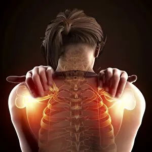
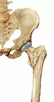
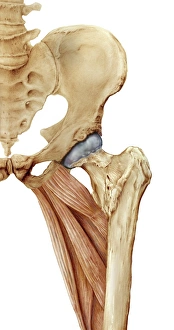
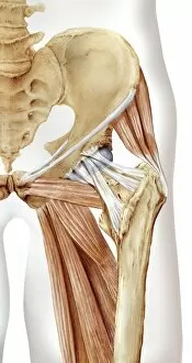
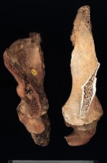
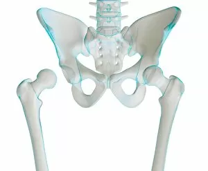
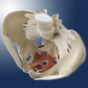
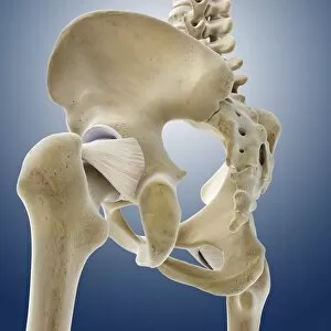
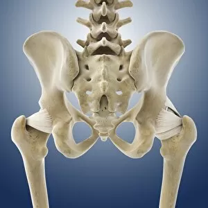
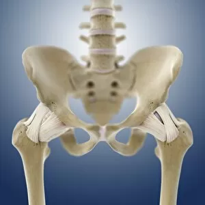
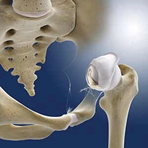
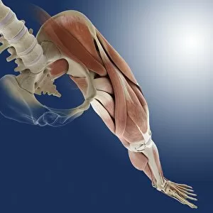
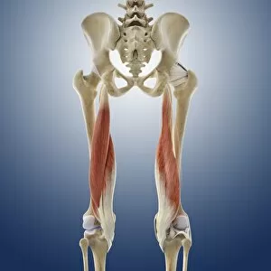
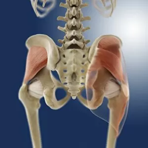
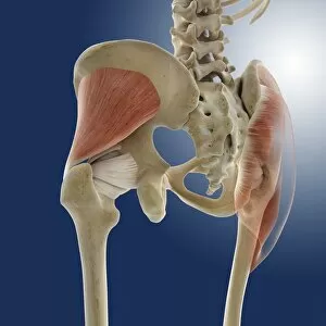
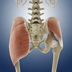
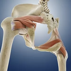
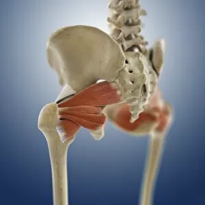
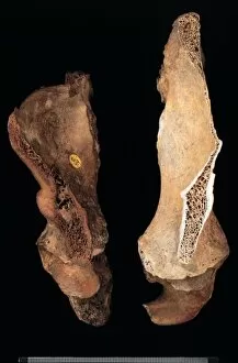
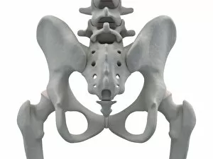
The ilium, a crucial part of the human pelvis, plays various roles in our body. In its normal anterior view, it forms the hip bones and provides support for the small intestine. However, repetitive activities like aerobics that involve lifting up the knee can lead to trauma in this area. Arthritis and osteophytes on femoral heads are visible when observing an anterior view of the pelvis with hip bones affected by these conditions. On the other hand, a normal open hip reveals the articular surface of the femur. Snapping hip syndrome occurs when there is subluxation of the iliopsoas tendon over the greater trochanter. This condition can cause discomfort during movement. Interestingly, historical manuscripts shed light on how Troy influenced French culture. For instance, "Ms 2028 fol. 2 The Trojan origins of the French nation" from Grandes Chroniques de depicts this connection while "Ms 782 The Construction of Troy" illustrates Le Roman de Troie. A posterior view showcases not only the hip bones but also includes a glimpse of gluteus maximus muscle and lumbar region anatomy. To better understand this intricate structure, studying an anterior view with labeled parts or examining male human skeleton views from both front and back angles proves beneficial in grasping its complexity.