
Immunofluorescent Collection
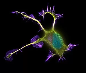
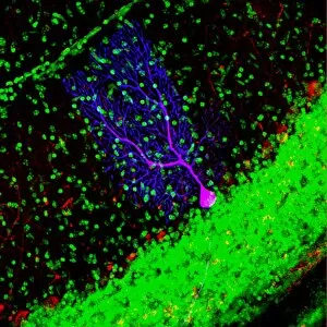
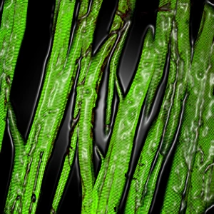
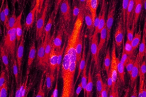
Immunofluorescent techniques revolutionize cell imaging, allowing us to delve into the intricate world of cellular structures and functions


All Professionally Made to Order for Quick Shipping
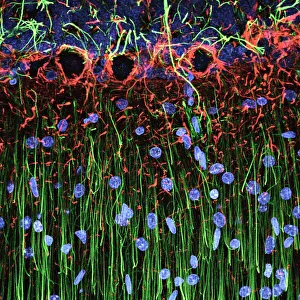
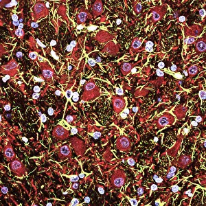
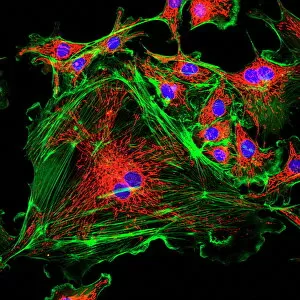
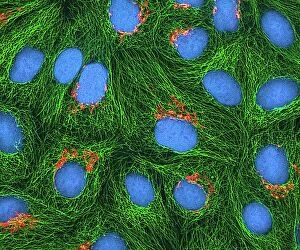
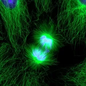
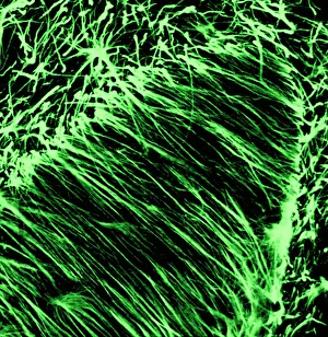
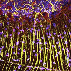
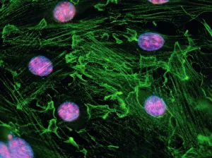
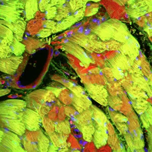
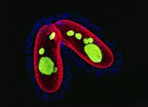
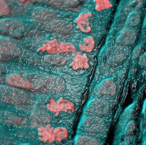
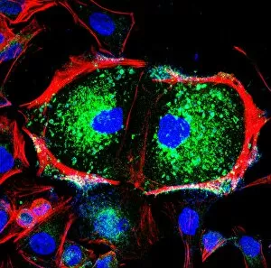
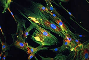
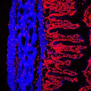
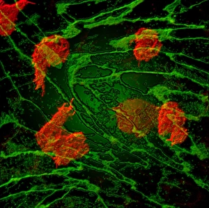
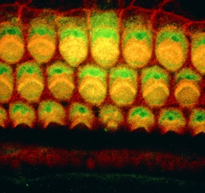
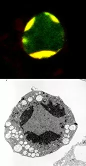
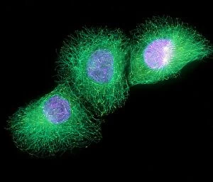
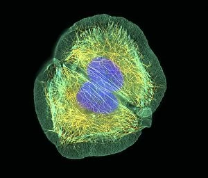
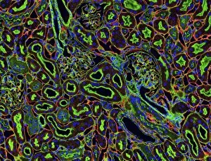
Immunofluorescent techniques revolutionize cell imaging, allowing us to delve into the intricate world of cellular structures and functions. In a stunning light micrograph of cerebellum tissue, we witness the vibrant fluorescence illuminating specific proteins or molecules within this brain region. This technique provides invaluable insights into the organization and communication between cells in this crucial part of our nervous system. HeLa cells, captured in another mesmerizing light micrograph (C017 / 8299), showcase their intricate cell structure highlighted by immunofluorescence. The fluorescent markers enable scientists to visualize various components within these immortalized human cells, unraveling their secrets one layer at a time. Witnessing mitosis through a captivating light micrograph unveils the beauty and complexity of cell division. Immunofluorescent staining allows us to track each stage with precision, shedding light on how our cells multiply and regenerate. Moving beyond HeLa cells (light micrograph C017 / 8298), we explore cerebral cortex nerve cells that play an integral role in cognition and sensory perception. Immunofluorescence unravels their distinct characteristics, enabling researchers to understand how they form connections essential for higher-order brain functions. Fluorescent micrographs capture uterine cells during childbirth—immunofluorescence reveals the dynamic changes occurring as these specialized muscle fibers contract rhythmically during labor. This visual insight deepens our understanding of reproductive physiology while highlighting the power techniques in studying complex biological processes. Returning once again to cerebellum tissue (light micrographs), we marvel at its intricate architecture revealed through immunofluorescence. By targeting specific proteins or molecules with fluorescent tags, scientists can decipher how different cell types interact within this vital brain region responsible for motor coordination and balance. Finally, an immunofluorescent LM image showcases fibroblast cell nuclei glowing brightly against a dark background—a testament to the precision and sensitivity of this technique.