
Lesions Collection
"Exploring the Intricate World of Lesions: From Boerhaave to Modern Medicine" Lesions, a term encompassing various types of tissue damage or abnormalities


All Professionally Made to Order for Quick Shipping
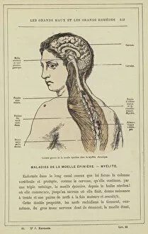
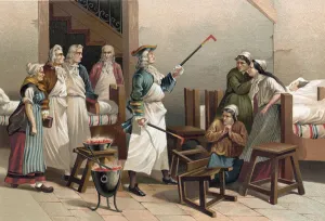
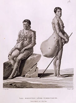
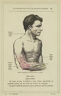
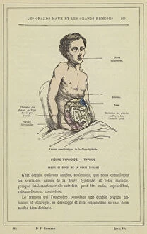
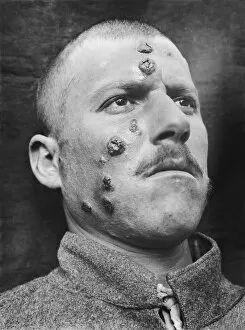
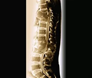
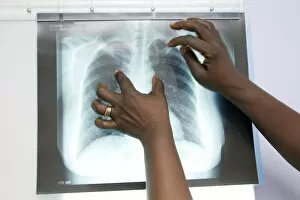
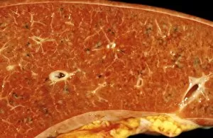
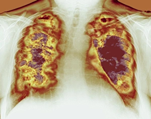
"Exploring the Intricate World of Lesions: From Boerhaave to Modern Medicine" Lesions, a term encompassing various types of tissue damage or abnormalities, have long fascinated medical professionals and researchers. One notable figure in the field was Herman Boerhaave, whose contributions revolutionized our understanding and their implications. Accidents can often lead to characteristic and significant lesions. The vividly depicted "Lesions caracteristiques et principales lesions du scorbut" lithograph showcases the effects of scurvy on the body. Similarly, "Lesions caracteristiques de la meningite tuberculeuse" lithograph highlights the distinct lesions associated with tuberculous meningitis. Chronic conditions like myelitis also leave profound marks on affected individuals. Through color lithographs such as "Lesions graves de la moelle epiniere dans la myelite chronique, " we gain insight into the severe spinal cord lesions seen in chronic myelitis patients. Boerhaave's groundbreaking work extended beyond lesion studies; he even delved into hysteric women's treatment at Leyden Hospital. His observations shed light on how certain psychological factors could manifest as physical symptoms and result in specific lesions. In different parts of the world, diseases like tuberculosis continue to afflict individuals today. A poignant example is seen in an image from Northern Mariana Islands depicting two men suffering from tuberculosis-related lesions—a reminder that these conditions persist despite medical advancements. Diabetes can also cause organic alterations and lesions during its advanced stages, as illustrated by another captivating color lithograph titled "Alterations et lesiones organiques a la derniere periode du diabete. " Pulmonary diseases are notorious for leaving distinctive marks within lung tissues. The vibrant lithograph called "Lesions caracteristiques de la phthisie pulmonaire" visually captures these characteristic pulmonary lesion patterns associated with tuberculosis.