Fraud Blocker Skip to main content

Macromolecule Collection (page 2)

Macromolecules, the building blocks of life, are at the forefront of scientific innovation

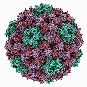
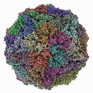
Background imageMacromolecule Collection: Cowpea chlorotic mottle virus capsid

Cowpea chlorotic mottle virus capsid, molecular model. This virus (CCMV) infects the cowpea plant (Vigna unguiculata), causing yellow spots of discolouration

Background imageMacromolecule Collection: Potassium ion channel protein structure

Potassium ion channel protein structure. Molecular model of a KcsA potassium ion (K+) channel from Streptomyces lividans bacteria

Background imageMacromolecule Collection: Streptavidin-biotin molecular complex

Streptavidin-biotin molecular complex. Molecular model of a single-strand binding complex of streptavidin (ribbons) and biotin (space-filled model, centre). Biotin is also known as vitamin B7

Background imageMacromolecule Collection: Potassium ion channel beta subunit

Potassium ion channel beta subunit. Molecular model showing the structure a beta subunit of a voltage-dependent potassium (K+) channel

Background imageMacromolecule Collection: KCNQ ion channel protein structure

KCNQ ion channel protein structure. Molecular model showing the protein structure of an ion channel domain. Ion channels are membrane-spanning proteins that form a pathway for the movement of
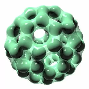
Background imageMacromolecule Collection: Potassium ion channel cavity structure

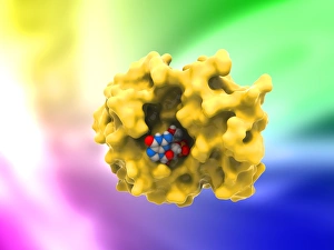
Potassium ion channel cavity structure. Molecular model showing the structure of a cavity formed by potassium ion channel proteins

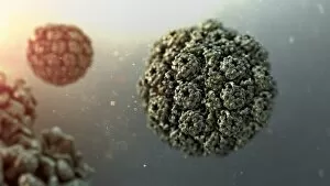
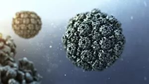
Background imageMacromolecule Collection: Avian polyomavirus capsid

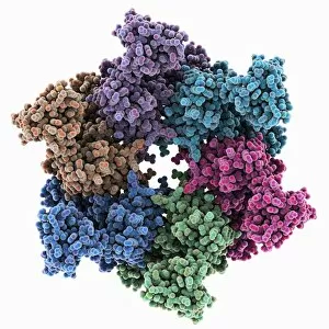
Avian polyomavirus capsid, molecular model. This virus, one of a range named for their potential to cause multiple tumours, infects birds. Discovered in budgerigars in 1981, it is often fatal

Background imageMacromolecule Collection: Cytoplasmic polyhedrosis virus capsid

Cytoplasmic polyhedrosis virus capsid, molecular model. Part of the Cypovirus genus and invariably fatal, this insect virus is transmitted by contamination of leaves eaten (examples include silkworms)

Background imageMacromolecule Collection: TATA box-binding protein complex C017 / 7090

TATA box-binding protein complex C017 / 7090
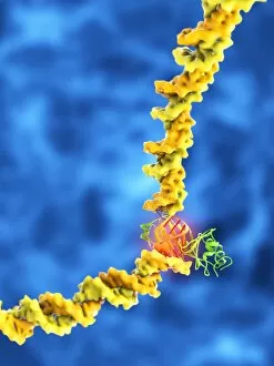
TATA box-binding protein complex. Molecular model showing a TATA box-binding protein (TBP, green) complexed with a strand of DNA (deoxyribonucleic acid, spheres) and transcription factor IIB

Background imageMacromolecule Collection: Theilers encephalomyelitis virus capsid

Theilers encephalomyelitis virus capsid, molecular model. This virus, which causes brain and spinal cord inflammation in mice, is used in research

Background imageMacromolecule Collection: TATA box-binding protein complex C017 / 7085

TATA box-binding protein complex C017 / 7085
TATA box-binding protein complex. Molecular model showing a TATA box-binding protein (TBP, green) complexed with a strand of DNA (deoxyribonucleic acid, yellow) and transcription factor IIB

Background imageMacromolecule Collection: Ricin A-chain, artwork C017 / 3654

Ricin A-chain, artwork C017 / 3654
Ricin A-chain. Computer artwork showing the enzymatically active A-chain from a molecule of the toxic protein ricin. Ricin comprises two entwined amino acid chains; A (seen here) and B (not shown)

Background imageMacromolecule Collection: Tobacco necrosis virus capsid

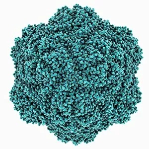
Tobacco necrosis virus capsid, molecular model. This plant virus infects a wide rage of plants, including the tobacco plant for which it is named. The virus causes tissue death (necrosis)

Background imageMacromolecule Collection: Ricin molecule, artwork C017 / 3649

Ricin molecule, artwork C017 / 3649
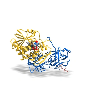
Ricin molecule. Computer artwork showing the structure of a molecule of the toxic protein ricin. Ricin comprises two entwined amino acid chains; A (yellow) and B (blue)
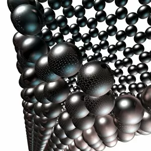
Background imageMacromolecule Collection: TATA box-binding protein complex C017 / 7083

TATA box-binding protein complex C017 / 7083
TATA box-binding protein complex. Molecular model showing a TATA box-binding protein (TBP, green) complexed with a strand of DNA (deoxyribonucleic acid, yellow) and transcription factor IIB

Background imageMacromolecule Collection: Grapevine fanleaf virus capsid

Grapevine fanleaf virus capsid, molecular model. This plant virus is named for its infection of grape vines. It is transmitted by the nematode worm Xiphinema index

Background imageMacromolecule Collection: VEE equine encephalitis virus capsid

VEE equine encephalitis virus capsid
Venezuelan equine encephalitis virus capsid, molecular model. This mosquito-borne virus can kill horses and other equine species, causing brain and spinal cord inflammation

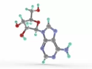
Background imageMacromolecule Collection: Adenosine molecule

Adenosine molecule
Adenosine monophosphate (AMP), molecular model. Nucleotide used as a monomer in RNA. Atoms are represented as spheres and are colour-coded: carbon (grey), hydrogen (green-blue), nitrogen (blue)

Background imageMacromolecule Collection: Murine minute virus capsid

Murine minute virus capsid, molecular model. This parvovirus infects mice, its only known natural host. It is highly infectious, transmitted through the nose and mouth

Background imageMacromolecule Collection: TATA box-binding protein complex C017 / 7089

TATA box-binding protein complex C017 / 7089
TATA box-binding protein complex. Molecular model showing a TATA box-binding protein (TBP, green) complexed with a strand of DNA (deoxyribonucleic acid, spheres) and transcription factor IIB

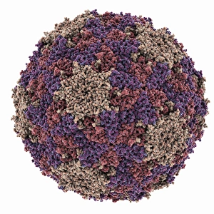
Background imageMacromolecule Collection: SV40 virus capsid, molecular model C018 / 7904

SV40 virus capsid, molecular model C018 / 7904
SV40 virus capsid, molecular model. Simian virus 40 (SV40) is found in monkeys such as Rhesus monkeys and macaques. Potentially tumour-causing, it is used in laboratory research and in vaccines

Background imageMacromolecule Collection: SV40 virus capsid, molecular model C018 / 7903

SV40 virus capsid, molecular model C018 / 7903
SV40 virus capsid, molecular model. Simian virus 40 (SV40) is found in monkeys such as Rhesus monkeys and macaques. Potentially tumour-causing, it is used in laboratory research and in vaccines

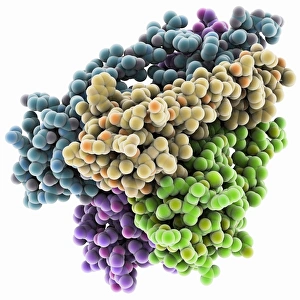
Background imageMacromolecule Collection: T-cell receptor bound to enterotoxin

T-cell receptor bound to enterotoxin, molecular model. The T cell receptor (TCR) is a protein complex found on the surface of a type of white blood cell called T lymphocytes (or T cells)

Background imageMacromolecule Collection: Bird egg white protein, molecular model

Bird egg white protein, molecular model. This is a deglycosylated form of the egg white glycoprotein avidin, obtained from a chicken (Gallus gallus)

Background imageMacromolecule Collection: Reversibly switchable fluorescent protein

Reversibly switchable fluorescent protein, molecular model. Reversibly switchable fluorescent proteins (RSFPs) are proteins that can be repeatedly converted between a fluorescent

Background imageMacromolecule Collection: Excisionase complex with DNA

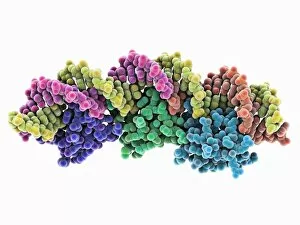
Excisionase complex with DNA. Molecular model of three excisionase proteins (bottom, purple, green and blue) bound to a strand of DNA (top, deoxyribonucleic acid)

Background imageMacromolecule Collection: Epstein-Barr virus protein and DNA

Epstein-Barr virus protein and DNA. Molecular model of the DNA-binding domain of a viral protein (pink-blue) bound to a lytic gene promoter element (viral strand of DNA, left)

Background imageMacromolecule Collection: DNA translocase, molecular model

DNA translocase, molecular model
ftsk, , protein, biomolecule, macromolecule, translocase, enzyme, pseudomonas aeruginosa, bacteria, biochemistry, biology, genetics, molecular biology, proteomics, artwork, illustration

Background imageMacromolecule Collection: Ebola virus transcription factor fragment

Ebola virus transcription factor fragment. Molecular model of the C-terminal domain (CTD) of Ebola virus transcription factor VP30

Background imageMacromolecule Collection: Bacterial twitching motility protein

Bacterial twitching motility protein
pilt, , protein, biomolecule, macromolecule, bacterial twitching motility, enzyme, aquifex aeolicus, bacterium, biochemistry, biology, molecular biology, proteomics, bacteriology, microbiology

Background imageMacromolecule Collection: RuvBL1 helicase enzyme

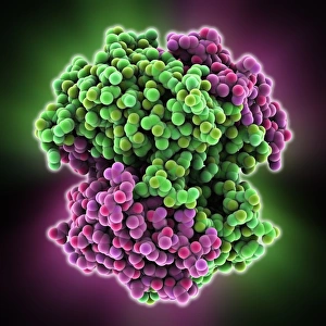
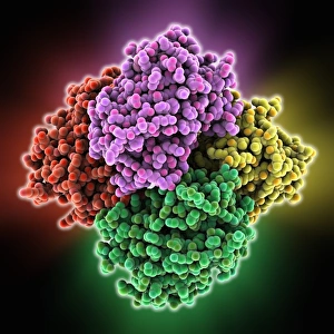
RuvBL1 helicase enzyme, molecular model. Helicases are enzymes that carry out several roles, primarily separating the two strands of the DNA (deoxyribonucleic acid) double helix

Background imageMacromolecule Collection: Metal-binding protein bound to DNA

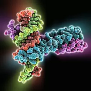
Metal-binding protein bound to DNA. Molecular model of the bacterial metal-binding protein NikR (bottom) bound to a strand of DNA (top, helical, deoxyribonucleic acid)

Background imageMacromolecule Collection: Muscle contraction proteins

Muscle contraction proteins. Molecular model of muscle protein motor cross-bridges during contraction in muscle. The cross-bridge is seen from the side, with contraction taking place horizontally

Background imageMacromolecule Collection: High-contrast direct DNA image, TEM

High-contrast direct DNA image, TEM
High-contrast direct DNA image. Coloured transmission electron micrograph (TEM) of the first high-contrast direct image of a bundle (fibre) of strands of DNA (deoxyribonucleic acid)

Background imageMacromolecule Collection: Buckminsterfullerene molecule C016 / 8354

Buckminsterfullerene molecule C016 / 8354
Buckminsterfullerene molecule. Computer artwork showing the molecular structure of buckminsterfullerene, a structurally distinct form (allotrope) of carbon that has 60 carbon atoms (black)

Background imageMacromolecule Collection: Nanotube structure, artwork C016 / 8888

Nanotube structure, artwork C016 / 8888
This image may not be used in educational posters Nanotube structure. Computer artwork of the structure of a cylindrical nanotube

Background imageMacromolecule Collection: Nanotube structure, artwork C016 / 8889

Nanotube structure, artwork C016 / 8889
Nanotube structure. Computer artwork of the structure of a cylindrical nanotube. This molecule is a type of fullerene, a structural type (allotrope) of carbon

Background imageMacromolecule Collection: Nanotube structure, artwork C016 / 8886

Nanotube structure, artwork C016 / 8886
Nanotube structure. Computer artwork of the structure of a cylindrical nanotube. This molecule is a type of fullerene, a structural type (allotrope) of carbon

Background imageMacromolecule Collection: Nanotube structure, artwork C016 / 8887

Nanotube structure, artwork C016 / 8887
Nanotube structure. Computer artwork of the structure of a cylindrical nanotube. This molecule is a type of fullerene, a structural type (allotrope) of carbon

Background imageMacromolecule Collection: Nanotube structure, artwork C016 / 8890

Nanotube structure, artwork C016 / 8890
Nanotube structure. Computer artwork of the structure of a cylindrical nanotube. This molecule is a type of fullerene, a structural type (allotrope) of carbon

Background imageMacromolecule Collection: Nanotube structure, artwork C016 / 8885

Nanotube structure, artwork C016 / 8885
Nanotube structure. Computer artwork of the structure of a cylindrical nanotube. This molecule is a type of fullerene, a structural type (allotrope) of carbon

Background imageMacromolecule Collection: Nanotube structure, artwork C016 / 8891

Nanotube structure, artwork C016 / 8891
Nanotube structure. Computer artwork of the structure of a cylindrical nanotube. This molecule is a type of fullerene, a structural type (allotrope) of carbon

Background imageMacromolecule Collection: Nanotube structure, artwork C016 / 8883

Nanotube structure, artwork C016 / 8883
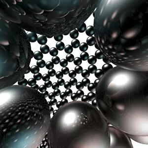
Nanotube structure. Computer artwork of the structure of a cylindrical nanotube. This molecule is a type of fullerene, a structural type (allotrope) of carbon

Background imageMacromolecule Collection: Nanotube structure, artwork C016 / 8884

Nanotube structure, artwork C016 / 8884
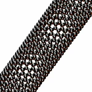
Nanotube structure. Computer artwork of the structure of a cylindrical nanotube. This molecule is a type of fullerene, a structural type (allotrope) of carbon

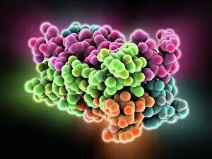
Background imageMacromolecule Collection: Alpha-beta T-cell receptor

Alpha-beta T-cell receptor, molecular model. The T cell receptor (TCR) is a protein complex found on the surface of a type of white blood cell called T lymphocytes (or T cells)

Background imageMacromolecule Collection: Carbamoylsarcosine amidase enzyme

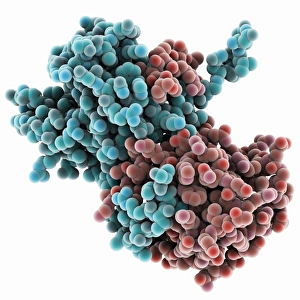
Carbamoylsarcosine amidase enzyme, molecular model. This enzyme catalyses the chemical reaction that converts the chemical N-carbamoylsarcosine in the presence of water to sarcosine (N-methylglycine)

Background imageMacromolecule Collection: Genomic HIV-RNA duplex

Genomic HIV-RNA duplex, molecular model. This structure shows the dimerization initiation site of genomic HIV-1 with RNA (ribonucleic acid)

Background imageMacromolecule Collection: Transducin protein beta-gamma complex

Transducin protein beta-gamma complex. Molecular model of the beta-gamma dimer of the heterotrimeric G protein transducin


All Professionally Made to Order for Quick Shipping

Macromolecules, the building blocks of life, are at the forefront of scientific innovation. Nanotube technology has revolutionized various fields, enabling advancements in medicine and electronics. In this captivating computer artwork, we witness the intricate Zinc fingers binding to a DNA strand, showcasing their crucial role in gene regulation. Carbon nanotubes have also emerged as remarkable materials with immense potential. Their unique structure and properties make them ideal for applications ranging from energy storage to drug delivery systems. Computer-generated images depict these carbon nanotubes in all their glory. The SARS coronavirus protein is another macromolecule that has garnered significant attention due to its role in viral infection. Scientists tirelessly study it to develop effective treatments against deadly outbreaks. Computer models allow us to explore complex structures like Bacteriophage phi29—a virus that infects bacteria—providing insights into its mechanisms and aiding in the development of targeted therapies. Simian immunodeficiency virus (SIV), closely related to HIV, poses a global health challenge. Understanding its macromolecular components helps researchers devise strategies for prevention and treatment. Rhodopsin protein molecule captures our imagination with its vital function in vision. Its elegant structure enables light detection and initiates visual signals within our eyes. TFAM transcription factor bound to DNA C015/7059 showcases how macromolecules regulate gene expression by interacting with specific regions on DNA strands—an essential process for cell functioning and development. These glimpses into the world of macromolecules highlight their significance across diverse disciplines—from cutting-edge technologies like nanotube engineering to unraveling infectious diseases or understanding fundamental biological processes. As scientists continue exploring these fascinating molecules, they pave the way for groundbreaking discoveries that shape our future.