
Meningitis Collection
Stephanie Lush, a courageous survivor of meningitis, defied all odds after having both her legs amputated due to this devastating disease


All Professionally Made to Order for Quick Shipping
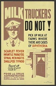
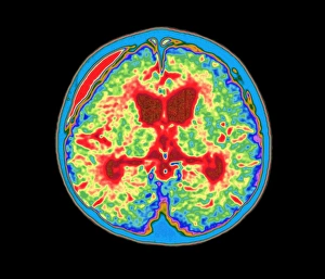
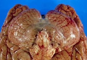
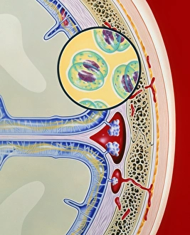
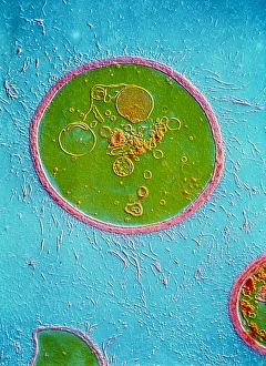
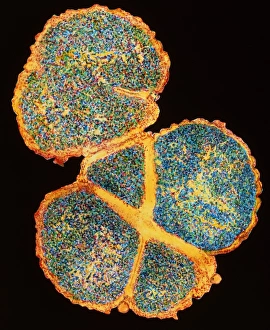
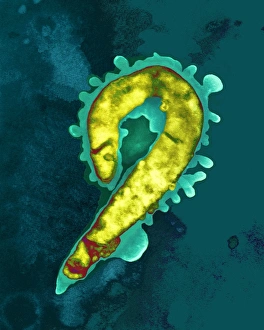
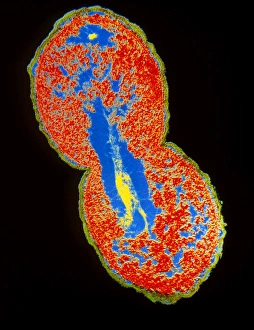
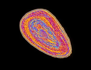
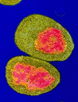
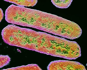
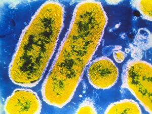
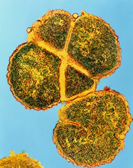
Stephanie Lush, a courageous survivor of meningitis, defied all odds after having both her legs amputated due to this devastating disease. Her inspiring journey serves as a reminder of the resilience and strength that can emerge from adversity. Lesions characteristic of tuberculous they can depicted in a vivid color lithograph, showcasing the visual impact this form of the disease can have on the human body. Such illustrations help medical professionals identify and treat patients more effectively. Nurses tirelessly stand by their patients' bedside during battles against meningitis, providing compassionate care and support. Their dedication plays an integral role in helping individuals like Stephanie regain their health and rebuild their lives. In 1872, mortality rates from smallpox and they were closely monitored in New York City. A contemporary lithograph chart correlated these rates with humidity and temperature fluctuations throughout the year, highlighting environmental factors that may influence the spread or severity of these diseases. Microscopic images reveal the menacing appearance bacteria under scanning electron microscopy (SEM). These visuals aid scientists in understanding its structure better for potential treatment development. The molecular model depicting Echovirus 7 capsid showcases intricate details at a microscopic level. Understanding such structures is crucial for researchers working towards developing effective vaccines or antiviral treatments against this specific strain of viral meningitis. During World War II, penicillin injections became vital tools in combating various infections, including those caused by bacterial strains associated with meningitis. This historical image represents one aspect of medical advancements made during times of crisis to save lives on battlefields around the world. Farmers take precautions to prevent milk contamination by ensuring milk truckers do not pick up milk where there is a risk of transmitting diseases like meningitis through infected animals or unsanitary conditions. These measures protect consumers from potential health risks associated with contaminated dairy products. Transmission electron microscopy (TEM) reveals detailed images capturing Coxsackie virus particles, which can cause meningitis.