
Meniscus Collection
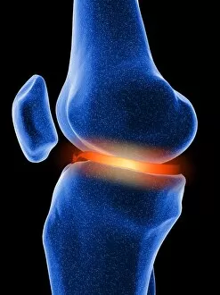
The meniscus, a crucial component of the anatomy of the human knee joint, plays a vital role in maintaining stability and preventing injury


All Professionally Made to Order for Quick Shipping
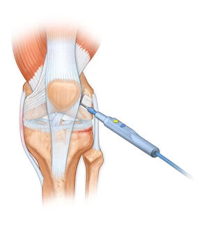
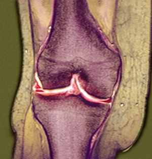
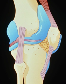
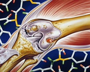
The meniscus, a crucial component of the anatomy of the human knee joint, plays a vital role in maintaining stability and preventing injury. During arthroscopic surgical procedures, a bovie is used to delicately cut through the retinaculum and clean up the femur of displaced patellar knees. An illustration of the anterior knee showcases the articular surface meniscus, highlighting its importance in facilitating smooth movement. Intriguingly, a camera obscura demonstrates how scenes outside are collected by a mirror tilted at 45 degrees, much like how our knowledge about meniscus has been gathered through meticulous observation and study. The insertion of arthroscopic instruments into the human knee reveals intricate details that aid surgeons during procedures. As we delve deeper into understanding this typical synovial joint, we uncover more about its complex structure and functions. Detailed illustrations provide insights into various arthroscopic surgical procedures performed on the knee. Amidst all these scientific explorations lies nature's beauty - a frog perched on a lily pad at a serene pond in Amador County, California. Just below the water's surface, another frog gracefully swims with its head pushing up against it; an enchanting sight from a front view perspective. Ultimately, it is through combining artistic depictions such as artwork F008 / 0073 with medical knowledge that we gain comprehensive insight into human knee anatomy. The multifaceted world continues to captivate us as we unravel its mysteries for both scientific advancement and appreciation of nature's wonders.