Knee anatomy, artwork C016 / 2883
![]()

Wall Art and Photo Gifts from Science Photo Library
Knee anatomy, artwork C016 / 2883
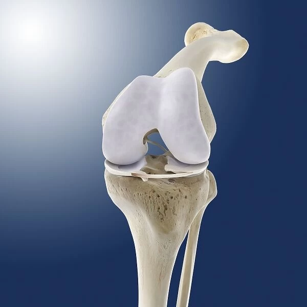
Knee flexion anatomy. Artwork of a frontal (anterior) view of the bones and some of the cartilage and ligaments of a flexed knee joint. The knee joint is where the upper leg bone (femur) articulates with the two lower leg bones, the tibia (left) and fibula (right). The ends of the bones are covered in pads of cartilage (white), with ligaments (white) holding the joint together. The ligament shown here is the transverse ligament, between the two menisci (lateral and medial) on the upper end of the tibia. The knee-cap (patella) is not shown
Science Photo Library features Science and Medical images including photos and illustrations
Media ID 9202707
© SPRINGER MEDIZIN/SCIENCE PHOTO LIBRARY
Anterior Arthrology Bent Cartilage Femur Fibula Flexed Flexion Frontal Hyaline Cartilage Joint Knee Ligament Ligaments Menisci Meniscus Osteology Tibia Blue Background Lateral Meniscus Medial Meniscus
EDITORS COMMENTS
This print showcases the intricate anatomy of a flexed knee joint. In this frontal (anterior) view, we can observe the bones and some of the cartilage and ligaments that make up this vital joint. The upper leg bone, known as the femur, articulates with two lower leg bones - the tibia on the left side and fibula on the right side. The ends of these bones are protected by pads of white cartilage, ensuring smooth movement within the joint. Ligaments play a crucial role in holding everything together, and one such ligament is prominently displayed here - it is called the transverse ligament located between two menisci: lateral and medial. Although not depicted in this artwork, it's important to note that there is also a knee-cap or patella present in our knees. This detailed illustration provides valuable insight into normal knee anatomy for both medical professionals and enthusiasts alike. With its blue background enhancing visual clarity, this image serves as an excellent resource for studying arthrology (the study of joints), osteology (the study of bones), and overall human body structure. Whether exploring biology or simply appreciating anatomical beauty, this print offers a fascinating glimpse into one of our most essential joints – healthy, functional, and ready to support us through every step we take.
MADE IN AUSTRALIA
Safe Shipping with 30 Day Money Back Guarantee
FREE PERSONALISATION*
We are proud to offer a range of customisation features including Personalised Captions, Color Filters and Picture Zoom Tools
SECURE PAYMENTS
We happily accept a wide range of payment options so you can pay for the things you need in the way that is most convenient for you
* Options may vary by product and licensing agreement. Zoomed Pictures can be adjusted in the Cart.

