Home > Popular Themes > Human Body
Knee bones and ligaments, artwork C016 / 7012

![]()
Wall Art and Photo Gifts from Science Photo Library
Knee bones and ligaments, artwork C016 / 7012
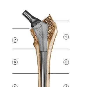
Knee bones and ligaments. Artwork of a frontal view of the knee joint, showing bones, ligaments, tendons, and cartilage. The bones are the femur (thigh bone, top), patella (knee-cap, upper centre), and the lower leg bones, the tibia (lower centre) and the fibula (lower right). The quadriceps femoris muscle tendon passes over the patella, becoming the patellar ligament of the tibia. At left and right are the medial and fibular collateral ligaments. Two short ligaments (centre) join femur and tibia: the posterior and anterior cruciate ligaments. The menisci (grey) are cartilage pads cushioning the joint. Posterior thigh muscles (hamstrings, red) are at upper left
Science Photo Library features Science and Medical images including photos and illustrations
Media ID 9245603
© D & L GRAPHICS / SCIENCE PHOTO LIBRARY
Anterior Anterior Cruciate Ligament Biceps Femoris Bones Cartilage Femur Fibula Frontal Hamstring Hamstrings Joint Knee Knee Cap Ligament Ligaments Lower Leg Menisci Meniscus Muscles Patella Patellar Ligament Semimembranosus Semitendinosus Tendon Tendons Thigh Bone Tibia Fibular Collateral Ligament Medial Collateral Ligament Posterior Cruciate Ligament
EDITORS COMMENTS
This artwork, titled "Knee Bones and Ligaments" offers a detailed frontal view of the intricate structures within the knee joint. The print showcases the essential components that enable smooth movement and stability in this crucial part of our body. At first glance, we are drawn to the prominent bones featured in this illustration. The femur, or thigh bone, takes center stage at the top, while just below it lies the patella, commonly known as the knee-cap. Completing the lower leg framework are two vital bones: the tibia positioned in the lower center and its companion on the right side, called fibula. The artist skillfully highlights various ligaments throughout this composition. On both sides of the knee joint stand tall and strong collateral ligaments – medial on one side and fibular on another – ensuring lateral stability during movement. Additionally, two short but significant cruciate ligaments connect femur to tibia: posterior and anterior cruciate ligaments. To provide cushioning support for these bony structures during motion, cartilage pads called menisci can be seen in a subtle grey shade within this artwork. These flexible pads act as shock absorbers between bones while maintaining proper alignment. Finally, we observe muscles surrounding this complex network of bones and ligaments. Posterior thigh muscles known as hamstrings grace us with their presence at upper left corner; they play a crucial role in bending our knees. Overall, this stunning visual representation serves as an educational tool for understanding human anatomy by showcasing healthy knee joints with utmost precision and detail.
MADE IN AUSTRALIA
Safe Shipping with 30 Day Money Back Guarantee
FREE PERSONALISATION*
We are proud to offer a range of customisation features including Personalised Captions, Color Filters and Picture Zoom Tools
SECURE PAYMENTS
We happily accept a wide range of payment options so you can pay for the things you need in the way that is most convenient for you
* Options may vary by product and licensing agreement. Zoomed Pictures can be adjusted in the Cart.