Fraud Blocker Skip to main content

Microbe Collection (page 3)

"Unveiling the Invisible: Exploring the Microbial World" In a realm unseen by the naked eye, lies a fascinating universe teeming with life

Background imageMicrobe Collection: Microscopy for the Million (litho)

Microscopy for the Million (litho)
7201243 Microscopy for the Million (litho) by Keene, Charles (1823-91); Private Collection; (add.info.: Microscopy for the million)

Background imageMicrobe Collection: Salt loving halobacteria turns a shallow lake bed red, Lone Pine, California, USA

Salt loving halobacteria turns a shallow lake bed red, Lone Pine, California, USA
Salt loving halobacteria turns a shallow salt lake bed red; Lone Pine, California, United States of America

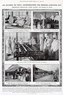
Background imageMicrobe Collection: Precautions taken against the plague in Japan, 1908

Precautions taken against the plague in Japan, 1908
An example to India: exterminating the microbe-carrying rat. Remarkable precautions taken against the plague in Japan. Photographs include sorting

Background imageMicrobe Collection: Leeuwenhoek the Microscope Man discovers the Microbe (litho)

Leeuwenhoek the Microscope Man discovers the Microbe (litho)
2812870 Leeuwenhoek the Microscope Man discovers the Microbe (litho) by English School, (20th century); Private Collection; (add.info.: Leeuwenhoek the Microscope Man discovers the Microbe)

Background imageMicrobe Collection: The Meeting of the British Association at Bath (engraving)

The Meeting of the British Association at Bath (engraving)
1041561 The Meeting of the British Association at Bath (engraving) by English School, (19th century); Private Collection; (add.info.: The Meeting of the British Association at Bath)

Background imageMicrobe Collection: Cover of 'The Butter Plate', number 477, Satirique en Colours

Cover of "The Butter Plate", number 477, Satirique en Colours
ICA4916826 Cover of " The Butter Plate", number 477, Satirique en Colours, 1910_5_21: Bread - Bread (mitron) - Illustration by Ricardo Flores (1878-1918) by Flores

Background imageMicrobe Collection: Alien wearing a face mask, illustration

Alien wearing a face mask, illustration

Background imageMicrobe Collection: ROBERT KOCH / IN KIMBERLEY

ROBERT KOCH / IN KIMBERLEY
HEINRICH HERMANN ROBERT KOCH German physician and pioneer bacteriologist in search of the Rinderpest microbe at Kimberley Date: 1843 - 1910

Background imageMicrobe Collection: Monster soup commonly called Thames water... 1828. Artist: Thomas McLean

Monster soup commonly called Thames water... 1828. Artist: Thomas McLean
Monster soup commonly called Thames water, being a correct representation of that precious stuff doled out to us!!!, 1828; showing an ugly woman holding a spy glass which touches the circumference of

Background imageMicrobe Collection: Microscopes and microscopical objects, 1750

Microscopes and microscopical objects, 1750. I: Wilsons pocket microscope. II: Scroll microscope. III: Tripod microscope - improved form of Marshalls double microscope

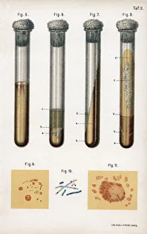
Background imageMicrobe Collection: Various types of bacteria, 1889

Various types of bacteria, 1889. 5: pneumonia culture; 6: albumen from a rotten egg; 7& 8: tuberculosis as prepared by Koch; 9: sputum from a TB patient; 10: anthrax bacillus; 11

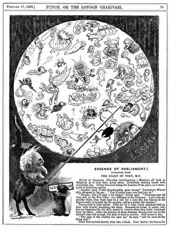
Background imageMicrobe Collection: Essence of Parliament, 1883. Artist: Harry Furniss

Essence of Parliament, 1883. Artist: Harry Furniss
Essence of Parliament, 1883. Cartoon marking the opening of the first session of Parliament for 1883, showing the politicians as microbes in a drop of water

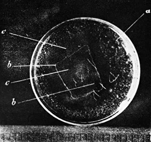
Background imageMicrobe Collection: Antibiotic action, historical image

Antibiotic action, historical image
Antibiotic action. Historical image taken by the German pathologist Karl Dohle (1855-1928) in 1889, showing the action of an antibiotic compound on Bacillus anthracis (anthrax)

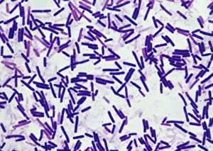
Background imageMicrobe Collection: Botulism bacteria

Botulism bacteria. Light micrograph of botulism bacteria (Clostridium botulinum). These bacteria occur naturally in soil. They produce botulinum toxin

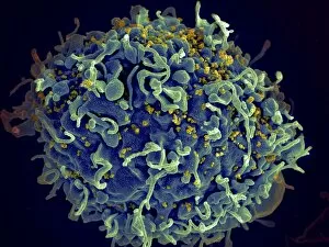
Background imageMicrobe Collection: Scanning electron micrograph of HIV particles infecting a human H9 T cell

Scanning electron micrograph of HIV particles infecting a human H9 T cell

Background imageMicrobe Collection: Coxiella burnetii, the bacteria that causes Q Fever

Coxiella burnetii, the bacteria that causes Q Fever. A dry fracture of a Vero cell exposing the contents of a vacuole where Coxiella burnetii are busy growing

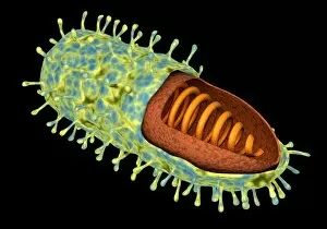
Background imageMicrobe Collection: Stylized rabies virus particles

Stylized rabies virus particles, the cause of the viral neuroinvasive disease acute encephalitis

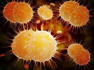
Background imageMicrobe Collection: Microscopic view of Neisseria gonorrhoeae

Microscopic view of Neisseria gonorrhoeae. Neisseria gonorrhoeae typically infects the mucous membranes causing infections such as urethritis, cervicitis, salpingitis, pelvic inflammatory disease

Background imageMicrobe Collection: Microscopic view of yellow fever virus

Microscopic view of yellow fever virus. Yellow fever is an acute viral disease

Background imageMicrobe Collection: Microscopic view of Giardiasis

Microscopic view of Giardiasis, an infectious disease caused by a unicellular parasite known as Giardia lamblia

Background imageMicrobe Collection: Microscopic view of bacterial pneumonia

Microscopic view of bacterial pneumonia. Bacterial pneumonia is a type of pneumonia caused by bacterial infection. Pneumonia can be generally defined as inflammation of the lung parenchyma

Background imageMicrobe Collection: Microscopic view of Streptococcus pneumoniae

Microscopic view of Streptococcus pneumoniae, also known as pneumococcus. Pneumococcus is a Gram-positive coccus shaped pathogenic bacteria which causes many types of pneumococcal infections in

Background imageMicrobe Collection: Conceptual image of rabies virus

Conceptual image of rabies virus

Background imageMicrobe Collection: Influenza virus particles, TEM

Influenza virus particles, TEM
Influenza virus particles, coloured transmission electron micrograph (TEM). The virus consists of ribonucleic acid (RNA), surrounded by a nucleocapsid (blue) and a lipid envelope (green)

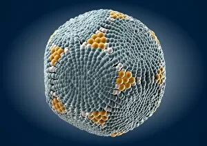
Background imageMicrobe Collection: Parvovirus particle, artwork C013 / 4640

Parvovirus particle, artwork C013 / 4640
Parvovirus particle. Computer artwork showing the outer structure of a human parvovirus (family Parvoviridae) particle (virion)

Background imageMicrobe Collection: Pyrococcus furiosus archaea artwork

Pyrococcus furiosus archaea artwork
Pyroccocus furiosus archaea, artwork. Archaea are single-celled organisms that are similar to bacteria but have been found to have higher organism characteristics

Background imageMicrobe Collection: Synthetic Mycoplasma bacteria, SEM C013 / 4777

Synthetic Mycoplasma bacteria, SEM C013 / 4777
Synthetic Mycoplasma bacteria. Coloured scanning electron micrograph (SEM) of Mycoplasma mycoides JCVI-syn1.0 cells. This was the first self-replicating bacterium controlled by a synthetic genome

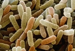
Background imageMicrobe Collection: Coloured SEM of Bacillus sp. bacteria

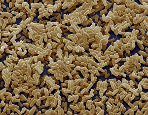
Coloured SEM of Bacillus sp. bacteria
Bacillus sp. Coloured scanning electron micrograph of bacteria cells (Bacillus sp.). These rod- shaped bacteria are found in soil and water and are usually saprotrophic

Background imageMicrobe Collection: Acetobacter and Schizosaccharomyces

Acetobacter and Schizosaccharomyces. Coloured scanning electron micrograph of Acetobacter bacteria and Schizosaccharomyces yeast cells in a culture of Kombucha" tea

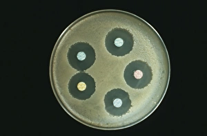
Background imageMicrobe Collection: Anthrax antibiotics research

Anthrax antibiotics research. Antibiotic drugs (discs) being tested on anthrax bacteria (Bacillus anthracis) colonies being cultured on an agar growth medium in a petri dish

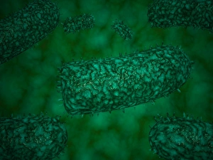
Background imageMicrobe Collection: Rod shaped bacillus bacteria

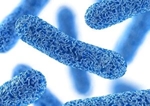
Rod shaped bacillus bacteria
3D-computer artwork of bacillus bacteria. These rod-shaped bacteria (bacilli) are widespread in soil and in the air. Many bacillus bacteria are responsible for food spoilage

Background imageMicrobe Collection: Infectious bronchitis virus (IBV), TEM

Infectious bronchitis virus (IBV), TEM
Infectious bronchitis virus (IBV) particles, coloured transmission electron micrograph (TEM). This virus is a member of the coronavirus family

Background imageMicrobe Collection: HIV particles, TEM

HIV particles, TEM
HIV (human immunodeficiency virus) particles, coloured transmission electron micrograph (TEM). Each particle consists of an RNA (ribonucleic acid, purple/brown) genome

Background imageMicrobe Collection: Bacterial lung infection

Bacterial lung infection, computer artwork. Rod-shaped bacteria (bacilli, green) on the surface of the alveoli of the lungs (purple)

Background imageMicrobe Collection: Mouldy bread

Mouldy bread. The white areas are fungal mycelia, masses of thread-like structures (hyphae) that absorb nutrients. The blue-green areas are the fungal spores (reproductive structures)

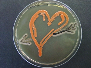
Background imageMicrobe Collection: Love, microbial art

Love, microbial art. Petri dish containing the fungi Aureobasidium pullulans and Rhodotorula sp. (red). The fungi have been cultured on agar jelly to depict a heart and arrow

Background imageMicrobe Collection: Bacillus subtilis bacteria, SEM

Bacillus subtilis bacteria, SEM
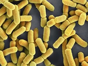
Bacillus subtilis. Coloured scanning electron micrograph (SEM) of Bacillus subtilis bacteria. B. subtilis, or hay bacillus, is an aerobic, gram-positive bacillus(rod-shaped bacterium)

Background imageMicrobe Collection: Lactobacillus bacteria, SEM

Lactobacillus bacteria, SEM
Lactobacillus sp. bacteria, coloured scanning electron micrograph (SEM). Lactobacillus bacteria are probiotic bacteria, which are useful for human and animal health as they produce bacteriocins

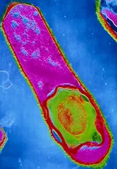
Background imageMicrobe Collection: Clostridium perfringens bacterium with spore

Clostridium perfringens bacterium with spore
False-colour transmission electron micrograph (TEM) of a Clostridium perfringens bacterium with endospore. This species of Gram-positive

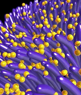
Background imageMicrobe Collection: Bacteria on lung hairs

Bacteria on lung hairs
Bacteria in a lung. Computer artwork of bacteria (yellow) on cilia (hair-like structures) in a lung. Bacterial infection of the lungs can cause potentially fatal pneumonia
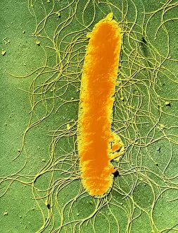
Background imageMicrobe Collection: Proteus mirabilis bacterium

Proteus mirabilis bacterium
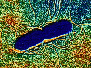
False-colour transmission electron micrograph of the bacterium Proteus mirabilis. It is an enterobacterium which is present normally in the human intestine
Background imageMicrobe Collection: Spirochete bacteria, TEM

Spirochete bacteria, TEM
Spirochete bacteria, coloured transmission electron micrograph (TEM). Spirochetes are bacteria with helically coiled cells

Background imageMicrobe Collection: Coloured SEM of Clostridium botulinum

Coloured SEM of Clostridium botulinum
Food poisoning bacterium. Coloured scanning electron micrograph of the Gram-positive anaerobic bacteria Clostridium botulinum, responsible for serious food poisoning in humans. C

Background imageMicrobe Collection: Rod-shaped bacteria

Rod-shaped bacteria. Computer artwork of rod- shaped bacteria (bacilli). Typical bacilli are E. coli and Salmonella bacteria, though there are many others as well

Background imageMicrobe Collection: Yeast cells, TEM

Yeast cells, TEM
Yeast cells, coloured transmission electron micrograph (TEM). The cell wall is shown in blue, cytoplasm in red, and vacuole in green. Yeast is a single-celled fungus that reproduces asexually

Background imageMicrobe Collection: Science / Leeuwenhoek

Science / Leeuwenhoek
ANTON VAN LEEUWENHOEK Dutch naturalist, depicted discovering the microbe Date: 1632 - 1723

Background imageMicrobe Collection: Illustration of immune response, involving chain of defensive white blood cells, triggered by microb

Illustration of immune response, involving chain of defensive white blood cells, triggered by microb

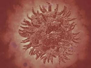
Background imageMicrobe Collection: Single virus particle

Single virus particle


All Professionally Made to Order for Quick Shipping

"Unveiling the Invisible: Exploring the Microbial World" In a realm unseen by the naked eye, lies a fascinating universe teeming with life. Meet the microbe, an extraordinary entity that encompasses various organisms and shapes our understanding of biology. HeLa cells, immortalized and widely used in research, have revolutionized medical science as they continue to unravel mysteries within our own bodies. Captured under a light microscope (C017 / 8299), their intricate structures reveal secrets waiting to be discovered. Witness the battle between neutrophils and MRSA as these microscopic warriors engage in a relentless struggle for survival. In an astonishing scanning electron microscope image (C018 / 8596), observe how these immune cells engulf harmful bacteria, showcasing nature's defense mechanisms at work. Eagerly multiplying like tiny soldiers on a mission, E. coli bacteria emerge into view through another SEM image. Their presence reminds us of both their beneficial role in digestion and potential harm when found in contaminated food or water sources. Salmonella bacteria take center stage next; their distinctive features magnified by yet another SEM image. These notorious culprits behind foodborne illnesses serve as reminders of proper hygiene practices necessary for safeguarding public health. Behold Yersinia pestis bacteria captured in vibrant colors through transmission electron microscopy (TEM). This captivating visual representation highlights the infamous pathogen responsible for devastating outbreaks such as the Black Death throughout history. Delving deeper into this microbial world reveals Candida fungus thriving amidst its surroundings. A striking SEM image showcases its filamentous structure – reminding us of its ability to cause infections if given favorable conditions. Returning to HeLa cells once more (light micrograph C017 / 8298), we are reminded of Henrietta Lacks' invaluable contribution to medical research - her immortal cell line continues to pave new paths towards scientific breakthroughs even decades after her passing.