Fraud Blocker Skip to main content

Microbe Collection (page 5)

"Unveiling the Invisible: Exploring the Microbial World" In a realm unseen by the naked eye, lies a fascinating universe teeming with life

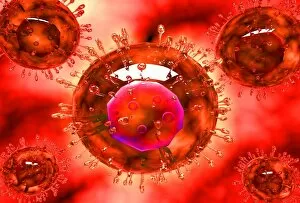
Background imageMicrobe Collection: Microscopic view of blood cells with virus

Microscopic view of blood cells with virus

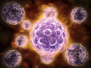
Background imageMicrobe Collection: Conceptual image of polyomavirus

Conceptual image of polyomavirus

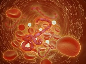
Background imageMicrobe Collection: Conceptual image of ebola virus in artery

Conceptual image of ebola virus in artery

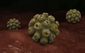
Background imageMicrobe Collection: Microscopic view of poliovirus

Microscopic view of poliovirus. Poliovirus is the causative agent of polio, a human enterovirus. It is composed of an RNA genome and a protein capsid

Background imageMicrobe Collection: Microscopic view of the capsid protein of Nudaurelia capensis omega virus

Microscopic view of the capsid protein of Nudaurelia capensis omega virus

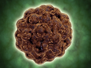
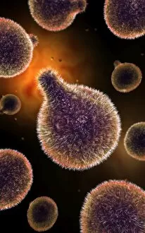
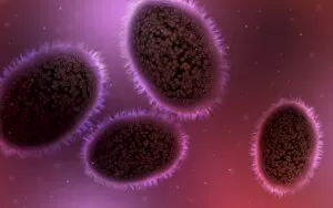
Background imageMicrobe Collection: Conceptual image of a viral spore

Conceptual image of a viral spore

Background imageMicrobe Collection: Microscopic view of Bacilli bacteria

Microscopic view of Bacilli bacteria

Background imageMicrobe Collection: Conceptual image of plasmodium causing malaria

Conceptual image of plasmodium causing malaria

Background imageMicrobe Collection: Microscopic view of Anthrax

Microscopic view of Anthrax. Anthrax is an acute disease caused by the bacterium Bacillus anthracis. Most forms of the disease are lethal, and it affects both humans and animals

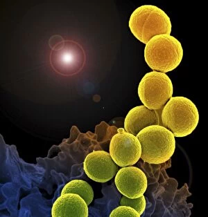
Background imageMicrobe Collection: Microscopic view of streptococcus

Microscopic view of streptococcus

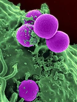
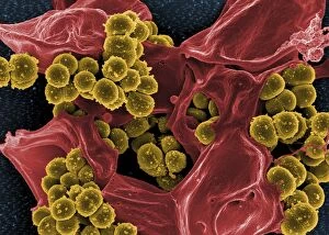
Background imageMicrobe Collection: A colorized scanning electron micrograph of a white blood cell eating MRSA

A colorized scanning electron micrograph of a white blood cell eating MRSA
A colorized scanning electron micrograph of a white blood cell eating an antibiotic resistant strain of Staphylococcus aureus bacteria, commonly known as MRSA

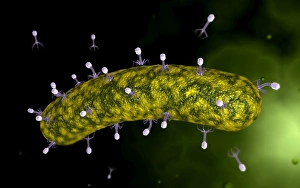
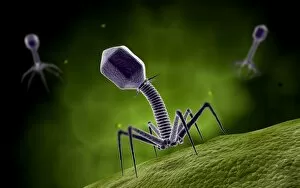
Background imageMicrobe Collection: Microscopic view of bacteriophage attacking bacteria

Microscopic view of bacteriophage attacking bacteria

Background imageMicrobe Collection: Conceptual image of Lactobacillus acidophilus

Conceptual image of Lactobacillus acidophilus. Lactobacillus is a gram-positive bacteria that occurs naturally in the human and animal gastrointestinal tract and mouth

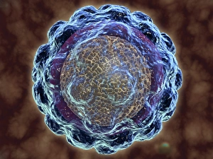
Background imageMicrobe Collection: Conceptual image of human cytomegalovirus

Conceptual image of human cytomegalovirus. Cytomegalovirus is a genus of the viral family Herpesviridae

Background imageMicrobe Collection: Microscopic view of Sindbis virus

Microscopic view of Sindbis virus (SINV). SINV is a mosquito-borne virus that causes rash and arthritis, has been causing outbreaks in humans

Background imageMicrobe Collection: Microscopic view of cocci bacterium

Microscopic view of cocci bacterium

Background imageMicrobe Collection: Conceptual image of salmonella typhi causing typhoid

Conceptual image of salmonella typhi causing typhoid

Background imageMicrobe Collection: Group of vibrio cholerae bacteria which causes cholera

Group of vibrio cholerae bacteria which causes cholera. Cholera is an infection in the small intestine caused by the bacterium Vibrio cholerae. The main symptoms are diarrhea and vomiting

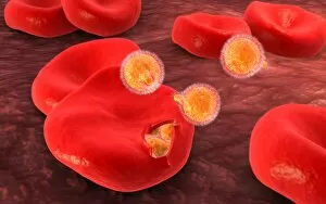
Background imageMicrobe Collection: Conceptual image of malaria parasites within red blood cells

Conceptual image of malaria parasites within red blood cells

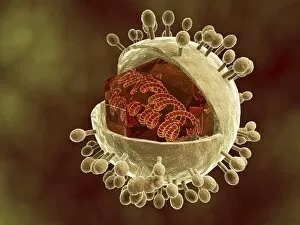
Background imageMicrobe Collection: Conceptual image of hepatitis virus

Conceptual image of hepatitis virus. Viral hepatitis is liver inflammation due to a viral infection. The most common causes of viral hepatitis are the five unrelated hepatotropic viruses

Background imageMicrobe Collection: Conceptual image of plasmodium

Conceptual image of plasmodium

Background imageMicrobe Collection: Scanning electron micrograph of a human neutrophil ingesting MRSA

Scanning electron micrograph of a human neutrophil ingesting MRSA

Background imageMicrobe Collection: Artistic representation of the immune systems reaction to bacteria invading the tissues

Artistic representation of the immune systems reaction to bacteria invading the tissues

Background imageMicrobe Collection: Microscopic view of samllpox

Microscopic view of samllpox

Background imageMicrobe Collection: Conceptual image of Euglena

Conceptual image of Euglena

Background imageMicrobe Collection: Group of H5N1 virus with glassy view

Group of H5N1 virus with glassy view

Background imageMicrobe Collection: Swine influenza virus

Swine influenza virus

Background imageMicrobe Collection: Microscopic view of Tetanus

Microscopic view of Tetanus. Tetanus is caused by the tetanus bacterium, Clostridium tetani. Tetanus is often associated with rust

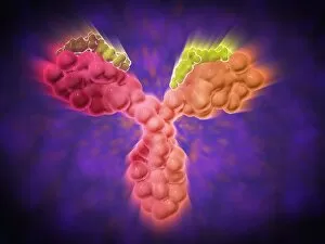
Background imageMicrobe Collection: Microscopic view of a human antibody

Microscopic view of a human antibody. Human antibodies are the Y-shaped proteins used by the immune system to defend against foreign objects like bacteria and viruses

Background imageMicrobe Collection: Conceptual image of the Zika virus

Conceptual image of the Zika virus

Background imageMicrobe Collection: Conceptual image of a tumor in human brain

Conceptual image of a tumor in human brain

Background imageMicrobe Collection: Conceptual image of Encephalitis

Conceptual image of Encephalitis. Encephalitis can cause flu-like symptoms, such as a fever or severe headaches. It can also cause confused thinking, seizures, or problems with senses

Background imageMicrobe Collection: Microscopic view of respiratory syncytial virus

Microscopic view of respiratory syncytial virus (RSV). RSV is a common virus that leads to mild, cold-like symptoms in adults and children

Background imageMicrobe Collection: Conceptual image of coxsackievirus

Conceptual image of coxsackievirus

Background imageMicrobe Collection: Microscopic view of Listeria monocytogenes

Microscopic view of Listeria monocytogenes. Listeria monocytogenes is the causative agent of the bacterial infection called listeriosis

Background imageMicrobe Collection: Scanning electron micrograph of Staphylococcus and a dead human neutrophil

Scanning electron micrograph of Staphylococcus and a dead human neutrophil
Scanning electron micrograph of methicillin-resistant Staphylococcus aureus and a dead human neutrophil

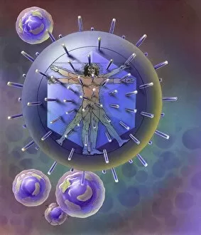
Background imageMicrobe Collection: Vitruvian Man inside virus particle

Vitruvian Man inside virus particle

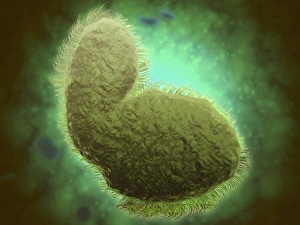
Background imageMicrobe Collection: Microscopic view of Paramecium bursaria

Microscopic view of Paramecium bursaria, a species of ciliate protozoan that has a mutualistic symbiotic relationship with green algae called Zoochlorella

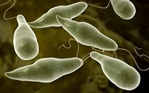
Background imageMicrobe Collection: Microscopic view of protozoa

Microscopic view of protozoa. Leishmaniasis and dysentery are examples of diseases involving protozoa, often transmitted to humans by the bite of a parasite-carrying insect

Background imageMicrobe Collection: Microscopic view of stem cell development

Microscopic view of stem cell development. In adult organisms, stem cells and progenitor cells act as a repair system for the body, replenishing adult tissues

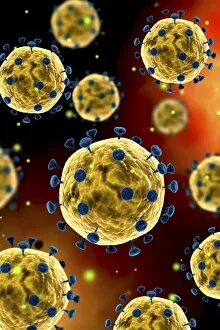
Background imageMicrobe Collection: Microscopic view of coronavirus

Microscopic view of coronavirus

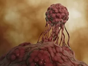
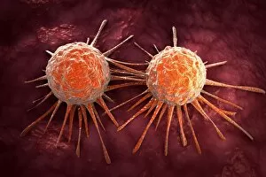
Background imageMicrobe Collection: Conceptual image of cancer virus

Conceptual image of cancer virus

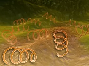
Background imageMicrobe Collection: Microscopic view of Syphillis

Microscopic view of Syphillis. Syphilis is a sexually transmitted infection caused by the spirochete bacterium, Treponema pallidum

Background imageMicrobe Collection: Microscopic view of athletes foot fungus

Microscopic view of athletes foot fungus. Athletes foot fungus, also known as ringworm of the foot and tinea pedis, is a fungal infection of the skin

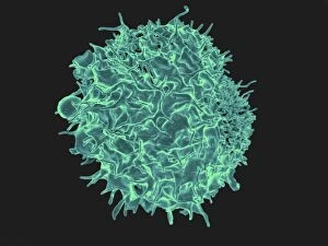
Background imageMicrobe Collection: Colorized scanning electron micrograph of a T lymphocyte

Colorized scanning electron micrograph of a T lymphocyte

Background imageMicrobe Collection: Microscopic view of bacteriophage

Microscopic view of bacteriophage

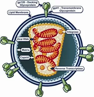
Background imageMicrobe Collection: Structure of HIV

Structure of HIV

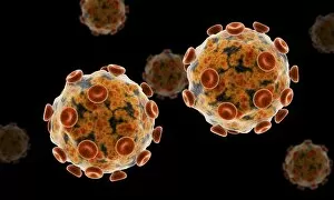
Background imageMicrobe Collection: Grouping of virus particles

Grouping of virus particles


All Professionally Made to Order for Quick Shipping

"Unveiling the Invisible: Exploring the Microbial World" In a realm unseen by the naked eye, lies a fascinating universe teeming with life. Meet the microbe, an extraordinary entity that encompasses various organisms and shapes our understanding of biology. HeLa cells, immortalized and widely used in research, have revolutionized medical science as they continue to unravel mysteries within our own bodies. Captured under a light microscope (C017 / 8299), their intricate structures reveal secrets waiting to be discovered. Witness the battle between neutrophils and MRSA as these microscopic warriors engage in a relentless struggle for survival. In an astonishing scanning electron microscope image (C018 / 8596), observe how these immune cells engulf harmful bacteria, showcasing nature's defense mechanisms at work. Eagerly multiplying like tiny soldiers on a mission, E. coli bacteria emerge into view through another SEM image. Their presence reminds us of both their beneficial role in digestion and potential harm when found in contaminated food or water sources. Salmonella bacteria take center stage next; their distinctive features magnified by yet another SEM image. These notorious culprits behind foodborne illnesses serve as reminders of proper hygiene practices necessary for safeguarding public health. Behold Yersinia pestis bacteria captured in vibrant colors through transmission electron microscopy (TEM). This captivating visual representation highlights the infamous pathogen responsible for devastating outbreaks such as the Black Death throughout history. Delving deeper into this microbial world reveals Candida fungus thriving amidst its surroundings. A striking SEM image showcases its filamentous structure – reminding us of its ability to cause infections if given favorable conditions. Returning to HeLa cells once more (light micrograph C017 / 8298), we are reminded of Henrietta Lacks' invaluable contribution to medical research - her immortal cell line continues to pave new paths towards scientific breakthroughs even decades after her passing.