
Microscopy Collection (page 7)
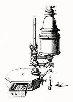
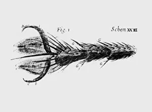
Microscopy, the art of exploring the hidden world beyond our naked eyes, unveils breathtaking wonders that captivate both scientists and artists alike


All Professionally Made to Order for Quick Shipping
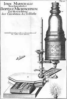
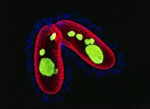
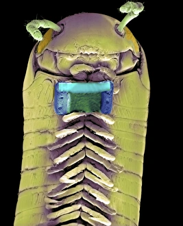
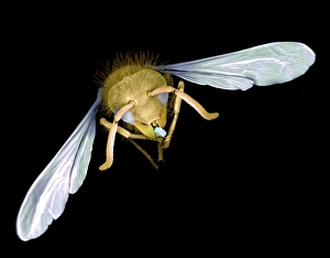
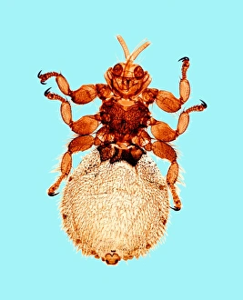
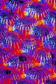
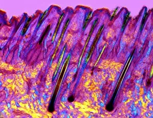
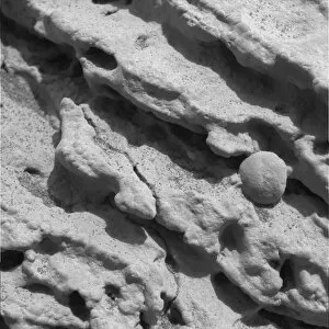
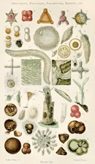
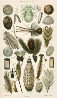
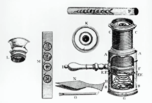
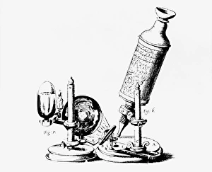
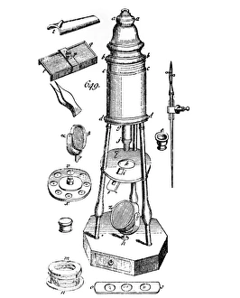
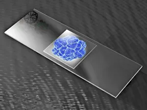
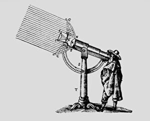
Microscopy, the art of exploring the hidden world beyond our naked eyes, unveils breathtaking wonders that captivate both scientists and artists alike. From intricate bee anatomy to historical artwork, each specimen under the microscope tells a unique story. In the depths of a laboratory, EDTA crystals shimmer like tiny diamonds as they reveal their molecular secrets through a mesmerizing light micrograph. Meanwhile, an illustration of a Flea C017/3435 transports us back in time, reminding us of the meticulous observations made by early pioneers. Snowflake research in the 18th century takes center stage as delicate ice crystals dance before our eyes. Pine pollen grains and pine stems come alive under the lens, showcasing their intricate structures with stunning clarity. The lime tree stem follows suit, revealing its hidden beauty through another enchanting light micrograph. Anton van Leeuwenhoek's groundbreaking discoveries come to life as we gaze upon his meticulously drawn animalcules from c1795. These microscopic organisms remind us of how far we have come in unraveling nature's mysteries. Water fleas and green algae form a delicate ecosystem within garden pond water—a harmonious dance captured for eternity by microscopy's watchful eye. Chlorophyte or green alga Volvox aureus floats gracefully in UK pond water—its larger spheres measuring just about 0—an awe-inspiring sight that reminds us of nature's infinite diversity. An abstract close-up reveals butterfly wing scales' vibrant colors and intricate patterns—a testament to evolution's artistic touch on even the tiniest creatures. And finally, false-colored SEM showcases a Tardigrade amongst moss from Peak District National Park—a minuscule creature measuring only approximately 0. 1 millimeters long but holding immense fascination for researchers worldwide.