Fraud Blocker Skip to main content

Molecules Collection (page 14)

"Molecules

Background imageMolecules Collection: Follicle-stimulating hormone complex C016 / 2794

Follicle-stimulating hormone complex C016 / 2794
Follicle-stimulating hormone (FSH) complex with receptor, molecular model. FSH helps to regulate human sexual development and reproductive processes. In females, it acts on follicles in the ovaries

Background imageMolecules Collection: RACK1 protein C016 / 2792

RACK1 protein C016 / 2792
RACK1 protein, molecular model. RACK1 stands for Receptor for Activated C Kinase 1. This protein is also known as guanine nucleotide-binding protein subunit beta-2-like 1 (GNB2L1)

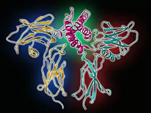
Background imageMolecules Collection: Follicle-stimulating hormone complex C016 / 2793

Follicle-stimulating hormone complex C016 / 2793
Follicle-stimulating hormone (FSH) complex with receptor, molecular model. FSH helps to regulate human sexual development and reproductive processes. In females, it acts on follicles in the ovaries

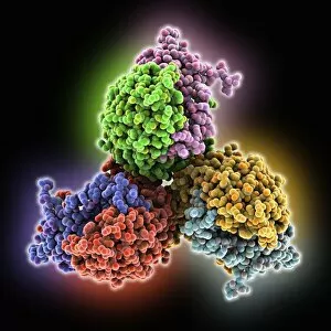
Background imageMolecules Collection: RACK1 protein C016 / 2791

RACK1 protein C016 / 2791
RACK1 protein, molecular model. RACK1 stands for Receptor for Activated C Kinase 1. This protein is also known as guanine nucleotide-binding protein subunit beta-2-like 1 (GNB2L1)

Background imageMolecules Collection: Human tumour protein p63 C016 / 2681

Human tumour protein p63 C016 / 2681
Human tumour protein p63. Molecular model of the tetramerization domain of the human tumour protein p63, encoded by the TP63 gene

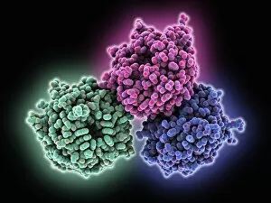
Background imageMolecules Collection: GMP synthetase enzyme C016 / 2679

GMP synthetase enzyme C016 / 2679
GMP synthetase enzyme, molecular model. This enzyme, guanine monophosphate synthetase, catalyses the reaction that converts xanthosine monophosphate to guanosine monophosphate

Background imageMolecules Collection: Human tumour protein p63 C016 / 2680

Human tumour protein p63 C016 / 2680
Human tumour protein p63. Molecular model of the tetramerization domain of the human tumour protein p63, encoded by the TP63 gene

Background imageMolecules Collection: GMP synthetase enzyme C016 / 2678

GMP synthetase enzyme C016 / 2678
GMP synthetase enzyme, molecular model. This enzyme, guanine monophosphate synthetase, catalyses the reaction that converts xanthosine monophosphate to guanosine monophosphate

Background imageMolecules Collection: Single-stranded DNA-binding protein C016 / 2676

Single-stranded DNA-binding protein C016 / 2676
Single-stranded DNA-binding protein, molecular model. The function of single-strand binding protein (SSB) is to bind to single strands of DNA (deoxyribonucleic acid) that form as DNA is replicated

Background imageMolecules Collection: Single-stranded DNA-binding protein C016 / 2675

Single-stranded DNA-binding protein C016 / 2675
Single-stranded DNA-binding protein, molecular model. The function of single-strand binding protein (SSB) is to bind to single strands of DNA (deoxyribonucleic acid) that form as DNA is replicated

Background imageMolecules Collection: RNA-silencing protein with RNA C016 / 2558

RNA-silencing protein with RNA C016 / 2558
RNA-silencing protein with RNA. Molecular model of RNA silencing taking place by human piwi-like protein (purple) acting on short loops (green) of RNA (ribonucleic acid)

Background imageMolecules Collection: RNA-silencing protein with RNA C016 / 2557

RNA-silencing protein with RNA C016 / 2557
RNA-silencing protein with RNA. Molecular model of RNA silencing taking place by human piwi-like protein (green) acting on short loops (red) of RNA (ribonucleic acid)

Background imageMolecules Collection: Plant anion channel protein homologue C016 / 2555

Plant anion channel protein homologue C016 / 2555
Plant anion channel protein homologue, molecular model. Obtained from the bacterium Haemophilus influenzae, this anion channel protein (TehA) is being studied due to its common structure (homologue)

Background imageMolecules Collection: Plant anion channel protein homologue C016 / 2556

Plant anion channel protein homologue C016 / 2556
Plant anion channel protein homologue, molecular model. Obtained from the bacterium Haemophilus influenzae, this anion channel protein (TehA) is being studied due to its common structure (homologue)

Background imageMolecules Collection: Sodium-potassium ion pump protein C016 / 2393

Sodium-potassium ion pump protein C016 / 2393
Sodium-potassium ion pump protein, molecular model. Sodium-potassium ATPase (adenosine triphosphatase) is an ATP-powered ion pump found in all animal cells

Background imageMolecules Collection: Sodium-potassium ion pump protein C016 / 2392

Sodium-potassium ion pump protein C016 / 2392
Sodium-potassium ion pump protein, molecular model. Sodium-potassium ATPase (adenosine triphosphatase) is an ATP-powered ion pump found in all animal cells

Background imageMolecules Collection: Dengue virus protein and antibody C016 / 2375

Dengue virus protein and antibody C016 / 2375
Dengue virus protein and antibody. Molecular model of a dengue virus 2 envelope protein (yellow) complexed with antibody Mab 4E11 (purple)

Background imageMolecules Collection: Helicase transcriptional silencer protein C016 / 2376

Helicase transcriptional silencer protein C016 / 2376
Helicase transcriptional silencer protein. Molecular model of the helicase protein MOM1 acting as a transcriptional silencer

Background imageMolecules Collection: Dengue virus protein and antibody C016 / 2374

Dengue virus protein and antibody C016 / 2374
Dengue virus protein and antibody. Molecular model of a dengue virus 2 envelope protein (red) complexed with antibody Mab 4E11 (blue)

Background imageMolecules Collection: Transthyretin blood protein C016 / 2327

Transthyretin blood protein C016 / 2327
Transthyretin blood protein. Molecular model of the thyroid hormone binding protein transthyretin, also known as prealbumin

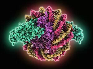
Background imageMolecules Collection: Sir3 gene silencer acting on DNA C016 / 2325

Sir3 gene silencer acting on DNA C016 / 2325
Sir3 gene silencer acting on DNA, molecular model. Sir3 (bright green) is acting on a circular strand of DNA (deoxyribonucleic acid, red and yellow)

Background imageMolecules Collection: Sir3 gene silencer acting on DNA C016 / 2324

Sir3 gene silencer acting on DNA C016 / 2324
Sir3 gene silencer acting on DNA, molecular model. Sir3 (light blue) is acting on a circular strand of DNA (deoxyribonucleic acid, pink)

Background imageMolecules Collection: Transthyretin blood protein C016 / 2326

Transthyretin blood protein C016 / 2326
Transthyretin blood protein. Molecular model of the thyroid hormone binding protein transthyretin, also known as prealbumin

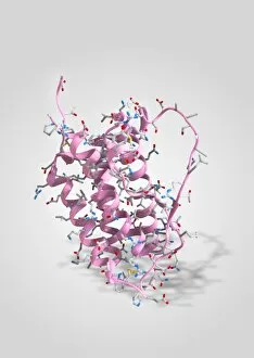
Background imageMolecules Collection: Neuropeptide Y neurotransmitter molecule C014 / 0013

Neuropeptide Y neurotransmitter molecule C014 / 0013
Neuropeptide Y neurotransmitter molecule. Molecular model showing the structure of the neurotransmitter neuropeptide Y (NPY)

Background imageMolecules Collection: Nano bearing, artwork C013 / 9992

Nano bearing, artwork C013 / 9992
Nano bearing, computer artwork. A bearing allows motion between two or more part. This bearing design is an example of nanotechnology

Background imageMolecules Collection: Human Apolipoprotein A-1 molecule

Human Apolipoprotein A-1 molecule. Molecular model showing the structure of a high-density lipoprotein (HDL) known as an apolipoprotein

Background imageMolecules Collection: DNA replication by helicase enzyme C013 / 9382

DNA replication by helicase enzyme C013 / 9382
Computer artwork of DNA Helicase breaking apart the hydrogen bonds of a DNA strand for replication. Helicases are a class of enzymes vital to all living organisms

Background imageMolecules Collection: Ribonuclease with RNA DNA hybrid

Ribonuclease with RNA DNA hybrid
Ribonuclease with RNA/DNA hybrid. Molecular model of Ribonuclease H (RNAse H, yellow and green) complexed with an RNA (ribonucleic acid, purple) and DNA (deoxyribonucleic acid, pink) hybrid

Background imageMolecules Collection: Cholesteryl ester transfer protein C013 / 8895

Cholesteryl ester transfer protein C013 / 8895
Cholesteryl ester transfer protein molecule. Computer model showing the structure of a molecule of cholesteryl ester transfer protein (CETP)

Background imageMolecules Collection: DNA Holliday junction complex C013 / 8888

DNA Holliday junction complex C013 / 8888
DNA Holliday junction complex. Molecular model of the enzyme FLP recombinase in complex with a Holliday junction between homologous strands of DNA (deoxyribonucleic acid)

Background imageMolecules Collection: Erythropoietin molecule C013 / 8891

Erythropoietin molecule C013 / 8891
Erythropoietin molecule (EPO), molecular model. EPO is a glycoprotein that is produced by the kidneys in response to low blood oxygen levels

Background imageMolecules Collection: Oxoguanine glycosylase complex C013 / 8886

Oxoguanine glycosylase complex C013 / 8886
Oxoguanine glycosylase complex. Computer model showing a molecule of human aG DNA repair glycosylase (right) bound to an DNA molecule (left)

Background imageMolecules Collection: Green fluorescent protein molecule C013 / 8885

Green fluorescent protein molecule C013 / 8885
Green fluorescent protein molecule. Computer model showing the secondary structure of a molecule of green fluorescent protein (GFP). GFP is found in the Pacific jellyfish Aequorea victoria

Background imageMolecules Collection: Oxoguanine glycosylase complex C013 / 8884

Oxoguanine glycosylase complex C013 / 8884
Oxoguanine glycosylase complex. Computer model showing an 8-Oxoguanine glycosylase (OGG1) molecule (green) bound to a section of DNA (deoxyribonucleic acid, pink and blue)

Background imageMolecules Collection: Erythropoietin hormone complex

Erythropoietin hormone complex. Computer model showing the secondary structure of a molecule of the human hormone Erythropoietin (EPO), complexed with an erythropoetin receptor molecule

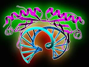
Background imageMolecules Collection: TATA box-binding protein complex C013 / 8881

TATA box-binding protein complex C013 / 8881
TATA box-binding protein complex. Molecular model showing a TATA box-binding protein (TBP) (purple) complexed with a strand of DNA (deoxyribonucleic acid, blue)

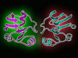
Background imageMolecules Collection: Bovine adrenodoxin molecules C013 / 8876

Bovine adrenodoxin molecules C013 / 8876
Bovine adrenodoxin molecules. Computer models showing the secondary structure of two molecules of adrenodoxin (adrenal ferredoxin) from a cow

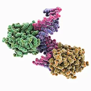
Background imageMolecules Collection: Pit-1 transcription factor bound to DNA C013 / 8872

Pit-1 transcription factor bound to DNA C013 / 8872
Pit-1 transcription factor bound to DNA. Molecular model showing pituitary-specific positive transcription factor 1 (Pit-1) (purple and yellow) bound to a strand of DNA (deoxyribonucleic acid)

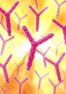
Background imageMolecules Collection: Antibodies, artwork C013 / 8800

Antibodies, artwork C013 / 8800
Antibodies. Computer artwork of antibody, or immunoglobulin, molecules. These y-shaped molecules have two arms that can bind to specific antigens, for instance viral or bacterial proteins

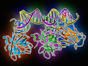
Background imageMolecules Collection: Tumour suppressor protein molecular model C013 / 7914

Tumour suppressor protein molecular model C013 / 7914
Tumour suppressor protein. Molecular model of the tumour suppressor protein p53 (blue, green and orange) bound to a molecule of DNA (deoxyribonucleic acid, yellow and pink)

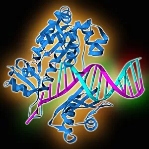
Background imageMolecules Collection: DNA polymerase molecule C013 / 7909

DNA polymerase molecule C013 / 7909
DNA polymerase. Molecular model of a molecule of DNA polymerase (blue) replicating a strand of DNA (deoxyribonucleic acid, pink and turquoise). The secondary structure of the DNA polymerase is shown

Background imageMolecules Collection: Antibodies, artwork C013 / 7792

Antibodies, artwork C013 / 7792
Antibodies. Computer artwork of antibody, or immunoglobulin, molecules. These y-shaped molecules have two arms that can bind to specific antigens, for instance viral or bacterial proteins

Background imageMolecules Collection: Antibodies, artwork C013 / 7791

Antibodies, artwork C013 / 7791
Antibodies. Computer artwork of antibody, or immunoglobulin, molecules. These y-shaped molecules have two arms that can bind to specific antigens, for instance viral or bacterial proteins

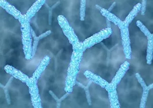
Background imageMolecules Collection: Antibodies, artwork C013 / 4683

Antibodies, artwork C013 / 4683
Antibodies. Computer artwork of antibody, or immunoglobulin, molecules. The y-shaped molecules have two arms that can bind to specific antigens, for instance viral or bacterial proteins

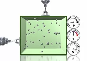
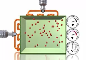
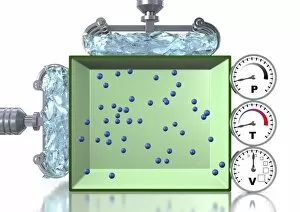
Background imageMolecules Collection: Boyles law of gases, artwork

Boyles law of gases, artwork
Boyles law of gases. Computer artwork explaining the gas laws as described by the combined gas law equation: PV=kT, which shows the relationship between the pressure (P), volume (V)

Background imageMolecules Collection: Combined gas law, artwork C013 / 4731

Combined gas law, artwork C013 / 4731
Combined gas law. Computer artwork explaining the gas laws as described by the combined gas law equation: PV=kT, which shows the relationship between the pressure (P), volume (V)

Background imageMolecules Collection: Pressure-temperature gas law, artwork C013 / 4730

Pressure-temperature gas law, artwork C013 / 4730
Pressure-temperature gas law. Computer artwork explaining the gas laws as described by the combined gas law equation: PV=kT, which shows the relationship between the pressure (P), volume (V)

Background imageMolecules Collection: Pressure-temperature gas law, artwork C013 / 4729

Pressure-temperature gas law, artwork C013 / 4729
Pressure-temperature gas law. Computer artwork explaining the gas laws as described by the combined gas law equation: PV=kT, which shows the relationship between the pressure (P), volume (V)


All Professionally Made to Order for Quick Shipping

"Molecules: The Building Blocks of Life and Beyond" From the intricate workings of an anaesthetic inhibiting an ion channel C015/6718 to the genius mind of James Clerk Maxwell, they have captivated scientists and artists alike. With their diverse structures and functions, they hold the key to understanding life at its core. Delving into the world of proteins, we witness their secondary structure through mesmerizing artwork that unveils their complexity. Meanwhile, the caffeine drug molecule keeps us awake while bacterial ribosomes tirelessly synthesize proteins within our cells. Vitamin B12's molecular model reminds us of nature's intricate design as zinc fingers elegantly bind to a DNA strand, orchestrating genetic processes. And who can forget capsaicin - the fiery molecule responsible for giving chili peppers their spicy kick? But molecules aren't limited to just earthly matters; they extend beyond our planet's boundaries. Oxytocin neurotransmitter molecules remind us of love's chemical connection while praziquantel parasite drugs combat infections in distant lands. Interferon molecules stand tall as defenders against viral invasions, showcasing our body's remarkable defense mechanisms. And amidst all this scientific wonder lies a breathtaking sight - Aurora Borealis dancing over a snow-covered coniferous forest in Northern Finland. Intricate and awe-inspiring, these glimpses into the molecular world remind us that there is so much more than meets the eye. From unlocking medical breakthroughs to unraveling nature's mysteries or simply marveling at captivating artistry – they can truly extraordinary entities shaping our understanding of life itself.