Fraud Blocker Skip to main content

Molecules Collection (page 4)

"Molecules

Background imageMolecules Collection: Transcription activation of IFN-beta gene F006 / 9510

Transcription activation of IFN-beta gene F006 / 9510
Transcription activation of IFN-beta gene. Molecular model of an enhanceosome containing the transcription factors IRF-3, ATF-2 and c-Jun bound to the interferon-beta (IFN-beta)
Background imageMolecules Collection: Transcription factor and DNA molecule F006 / 9484

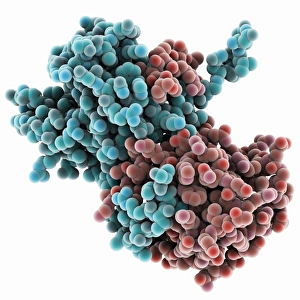
Transcription factor and DNA molecule F006 / 9484
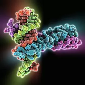
Transcription factor and DNA molecule. Molecular model of glucocorticoid receptor (GR) transcription factor protein (pink and blue) complexed with a molecule of DNA (deoxyribonucleic acid)

Background imageMolecules Collection: Haemagglutinin protein subunit F006 / 9479

Haemagglutinin protein subunit F006 / 9479
Haemagglutinin protein subunit. Molecular model of the ectodomain of the haemagglutinin HA(2) subunit. Haemagglutinin is a surface protein from the influenza A virus

Background imageMolecules Collection: Haemagglutinin viral surface protein F006 / 9470

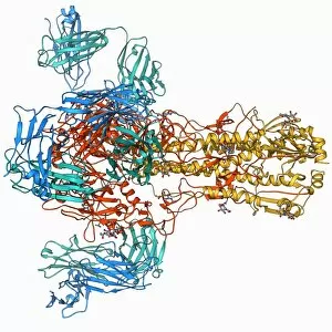
Haemagglutinin viral surface protein F006 / 9470
Haemagglutinin viral surface protein. Molecular model of haemagglutinin, a surface protein from the influenza virus, complexed with a neutralising antibody

Background imageMolecules Collection: ATPase and inhibitor, molecular model F006 / 9448

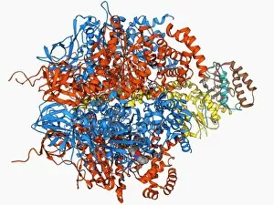
ATPase and inhibitor, molecular model F006 / 9448
ATPase and inhibitor. Computer model of an ATP synthase (ATPase) molecule from a mitochondrion complexed with its inhibitor protein IF1

Background imageMolecules Collection: Green fluorescent protein molecule F006 / 9402

Green fluorescent protein molecule F006 / 9402
Green fluorescent protein (GFP), molecular model. The molecule has a cylindrical structure formed from beta sheets (ribbons). GFP is found in the Pacific jellyfish Aequorea victoria

Background imageMolecules Collection: Green fluorescent protein molecule F006 / 9343

Green fluorescent protein molecule F006 / 9343
Green fluorescent protein (GFP), molecular model. The molecule has a cylindrical structure formed from beta sheets (ribbons). GFP is found in the Pacific jellyfish Aequorea victoria

Background imageMolecules Collection: Valyl-tRNA synthetase molecule F006 / 9342

Valyl-tRNA synthetase molecule F006 / 9342
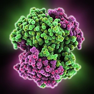
Valyl-tRNA synthetase protein molecule. Molecular model showing bacterial valyl-tRNA synthetase complexed with valyl tRNA (transfer ribonucleic acid)

Background imageMolecules Collection: DNA Holliday junction complex F006 / 9334

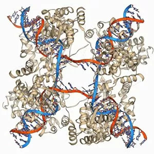
DNA Holliday junction complex F006 / 9334
DNA Holliday junction complex. Molecular model of the enzyme FLP recombinase in complex with a Holliday junction between homologous strands of DNA (deoxyribonucleic acid)

Background imageMolecules Collection: Isoleucyl-tRNA synthetase molecule F006 / 9329

Isoleucyl-tRNA synthetase molecule F006 / 9329
Isoleucyl-tRNA synthetase protein molecule. Molecular model showing bacterial isoleucyl-tRNA synthetase complexed with aspartyl tRNA (transfer ribonucleic acid)

Background imageMolecules Collection: Oxoguanine glycosylase complex F006 / 9318

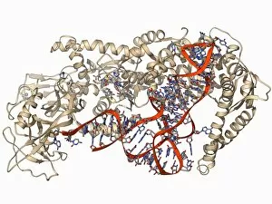
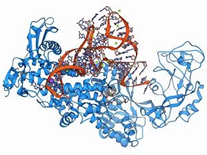
Oxoguanine glycosylase complex F006 / 9318
Oxoguanine glycosylase complex. Computer model showing an 8-Oxoguanine glycosylase (OGG1) molecule (beige) bound to a section of DNA (deoxyribonucleic acid, red and blue)

Background imageMolecules Collection: Restriction enzyme and DNA F006 / 9315

Restriction enzyme and DNA F006 / 9315
Restriction enzyme and DNA. Molecular model showing an EcoRI endonuclease enzyme (purple and green) bound to a DNA (deoxyribonucleic acid) molecule (red and blue)

Background imageMolecules Collection: Green fluorescent protein molecule F006 / 9313

Green fluorescent protein molecule F006 / 9313
Green fluorescent protein (GFP), molecular model. The molecule has a cylindrical structure formed from beta sheets (ribbons). GFP is found in the Pacific jellyfish Aequorea victoria

Background imageMolecules Collection: Oxoguanine glycosylase complex F006 / 9307

Oxoguanine glycosylase complex F006 / 9307
Oxoguanine glycosylase complex. Computer model showing an 8-Oxoguanine glycosylase (OGG1) molecule (beige) bound to a section of DNA (deoxyribonucleic acid, red and blue)

Background imageMolecules Collection: ATPase molecule F006 / 9300

ATPase molecule F006 / 9300
ATPase molecule. Molecular model of the central stalk of an ATP synthase (ATPase) molecule from a cow. ATPase is an important enzyme that provides energy for cells through the synthesis of adenosine

Background imageMolecules Collection: TATA box-binding protein and DNA F006 / 9267

TATA box-binding protein and DNA F006 / 9267
TATA box-binding protein and DNA. Molecular model showing a TATA box-binding protein (TBP) complexed with a strand of DNA (deoxyribonucleic acid, red and blue)

Background imageMolecules Collection: E coli Holliday junction complex F006 / 9261

E coli Holliday junction complex F006 / 9261
E. coli Holliday junction complex. Molecular model of a RuvA protein (red) in complex with a Holliday junction between homologous strands of DNA (deoxyribonucleic acid, blue) from an E

Background imageMolecules Collection: ATP synthase molecule F006 / 9258

ATP synthase molecule F006 / 9258
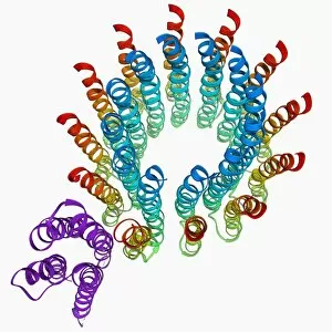
ATP synthase molecule. Molecular model showing the structure of ATP synthase (ATPase) subunit A and C. ATPase is an important enzyme that provides energy for cells through the synthesis of adenosine

Background imageMolecules Collection: Chaperonin protein complex F006 / 9236

Chaperonin protein complex F006 / 9236
Chaperonin protein complex. Molecular model showing the structure of a GroEL GroES (ADP)7 chaperonin complex. Chaperonins are proteins that provide favourable conditions for the correct folding of

Background imageMolecules Collection: Aspartyl-tRNA synthetase molecule F006 / 9238

Aspartyl-tRNA synthetase molecule F006 / 9238
Aspartyl-tRNA synthetase protein molecule. Molecular model showing bacterial aspartyl-tRNA synthetase complexed with aspartyl tRNA (transfer ribonucleic acid)

Background imageMolecules Collection: Interleukin-6, molecular model F006 / 9232

Interleukin-6, molecular model F006 / 9232
Interleukin-6. Molecular model of the cytokine protein human interleukin-6. This protein is produced in the body and has a wide variety of functions in the immune system

Background imageMolecules Collection: TATA box-binding protein complex F006 / 9230

TATA box-binding protein complex F006 / 9230
TATA box-binding protein complex. Molecular model showing a yeast TATA box-binding protein (TBP) complexed with a strand of DNA (deoxyribonucleic acid, red and blue) and transcription factor IIB

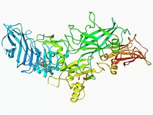
Background imageMolecules Collection: Anthrax protective antigen molecule F006 / 9229

Anthrax protective antigen molecule F006 / 9229
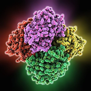
Anthrax protective antigen molecule. Computer model showing the structure of a molecule of protective antigen (PA) produced by anthrax (Bacillus anthracis) bacteria

Background imageMolecules Collection: Anthrax protective antigen molecule F006 / 9225

Anthrax protective antigen molecule F006 / 9225
Anthrax protective antigen molecule. Computer model showing the structure of a molecule of protective antigen (PA) produced by anthrax (Bacillus anthracis) bacteria

Background imageMolecules Collection: Rotaxane, molecular crystal structure C017 / 7016

Rotaxane, molecular crystal structure C017 / 7016
Molecular crystal structure of a rotaxane. A rotaxane is a chemical compound composed of a linear molecular chain passing through a chainlike molecular ring

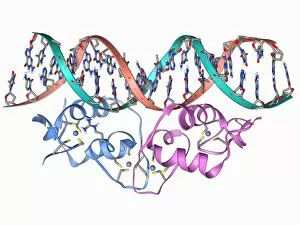
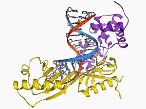
Background imageMolecules Collection: Zinc finger bound to DNA

Zinc finger bound to DNA. Molecular model showing a zinc finger molecule bound (orange) to a strand of DNA (deoxyribonucleic acid, pink and green)

Background imageMolecules Collection: TATA box-binding protein complex C017 / 7089

TATA box-binding protein complex C017 / 7089
TATA box-binding protein complex. Molecular model showing a TATA box-binding protein (TBP, green) complexed with a strand of DNA (deoxyribonucleic acid, spheres) and transcription factor IIB

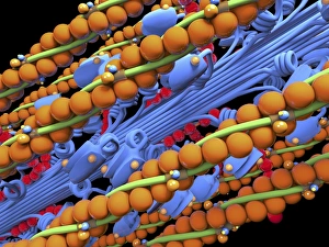
Background imageMolecules Collection: Actin Myosin Muscle Model, artwork C014 / 2661

Actin Myosin Muscle Model, artwork C014 / 2661
Computer artwork of the molecular actin myosin muscle structure. The complex ultrastructure of cells, their shape and internal structure

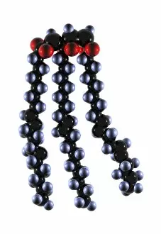
Background imageMolecules Collection: Polyunsaturated fat molecule

Polyunsaturated fat molecule. Computer model showing the structure of three unsaturated fat molecules bound together to form a polyunsaturated fat molecule

Background imageMolecules Collection: T-cell receptor bound to enterotoxin

T-cell receptor bound to enterotoxin, molecular model. The T cell receptor (TCR) is a protein complex found on the surface of a type of white blood cell called T lymphocytes (or T cells)

Background imageMolecules Collection: Bird egg white protein, molecular model

Bird egg white protein, molecular model. This is a deglycosylated form of the egg white glycoprotein avidin, obtained from a chicken (Gallus gallus)

Background imageMolecules Collection: Reversibly switchable fluorescent protein

Reversibly switchable fluorescent protein, molecular model. Reversibly switchable fluorescent proteins (RSFPs) are proteins that can be repeatedly converted between a fluorescent

Background imageMolecules Collection: Excisionase complex with DNA

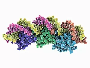
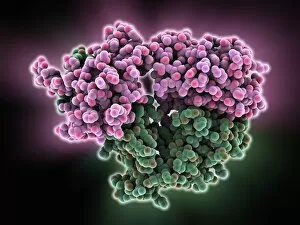
Excisionase complex with DNA. Molecular model of three excisionase proteins (bottom, purple, green and blue) bound to a strand of DNA (top, deoxyribonucleic acid)

Background imageMolecules Collection: Epstein-Barr virus protein and DNA

Epstein-Barr virus protein and DNA. Molecular model of the DNA-binding domain of a viral protein (pink-blue) bound to a lytic gene promoter element (viral strand of DNA, left)

Background imageMolecules Collection: DNA translocase, molecular model

DNA translocase, molecular model
ftsk, , protein, biomolecule, macromolecule, translocase, enzyme, pseudomonas aeruginosa, bacteria, biochemistry, biology, genetics, molecular biology, proteomics, artwork, illustration

Background imageMolecules Collection: Ebola virus transcription factor fragment

Ebola virus transcription factor fragment. Molecular model of the C-terminal domain (CTD) of Ebola virus transcription factor VP30

Background imageMolecules Collection: Bacterial twitching motility protein

Bacterial twitching motility protein
pilt, , protein, biomolecule, macromolecule, bacterial twitching motility, enzyme, aquifex aeolicus, bacterium, biochemistry, biology, molecular biology, proteomics, bacteriology, microbiology

Background imageMolecules Collection: RuvBL1 helicase enzyme

RuvBL1 helicase enzyme, molecular model. Helicases are enzymes that carry out several roles, primarily separating the two strands of the DNA (deoxyribonucleic acid) double helix

Background imageMolecules Collection: Metal-binding protein bound to DNA

Metal-binding protein bound to DNA. Molecular model of the bacterial metal-binding protein NikR (bottom) bound to a strand of DNA (top, helical, deoxyribonucleic acid)

Background imageMolecules Collection: Muscle contraction proteins

Muscle contraction proteins. Molecular model of muscle protein motor cross-bridges during contraction in muscle. The cross-bridge is seen from the side, with contraction taking place horizontally

Background imageMolecules Collection: Buckminsterfullerene molecule C016 / 8354

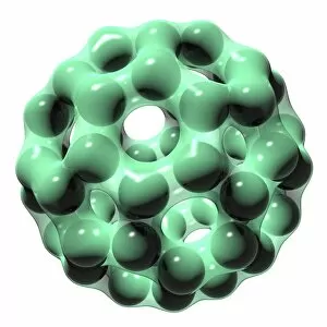
Buckminsterfullerene molecule C016 / 8354
Buckminsterfullerene molecule. Computer artwork showing the molecular structure of buckminsterfullerene, a structurally distinct form (allotrope) of carbon that has 60 carbon atoms (black)

Background imageMolecules Collection: Molecular structure, conceptual artwork C016 / 7534

Molecular structure, conceptual artwork C016 / 7534
Molecular structure, conceptual computer artwork

Background imageMolecules Collection: Alpha-beta T-cell receptor

Alpha-beta T-cell receptor, molecular model. The T cell receptor (TCR) is a protein complex found on the surface of a type of white blood cell called T lymphocytes (or T cells)

Background imageMolecules Collection: Carbamoylsarcosine amidase enzyme

Carbamoylsarcosine amidase enzyme, molecular model. This enzyme catalyses the chemical reaction that converts the chemical N-carbamoylsarcosine in the presence of water to sarcosine (N-methylglycine)

Background imageMolecules Collection: Genomic HIV-RNA duplex

Genomic HIV-RNA duplex, molecular model. This structure shows the dimerization initiation site of genomic HIV-1 with RNA (ribonucleic acid)

Background imageMolecules Collection: Transducin protein beta-gamma complex

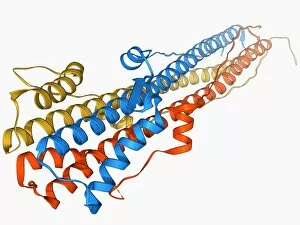
Transducin protein beta-gamma complex. Molecular model of the beta-gamma dimer of the heterotrimeric G protein transducin

Background imageMolecules Collection: Nerve growth factor bound to receptor

Nerve growth factor bound to receptor, molecular model. Nerve growth factor (NGF) complexed with the TrkA receptor. NGF is a neurotrophin that acts on the development and function of nerves

Background imageMolecules Collection: Chromosome segregation protein

Chromosome segregation protein, molecular model. This proteins function is to aid the process of chromosome segregation during cell division and replication


All Professionally Made to Order for Quick Shipping

"Molecules: The Building Blocks of Life and Beyond" From the intricate workings of an anaesthetic inhibiting an ion channel C015/6718 to the genius mind of James Clerk Maxwell, they have captivated scientists and artists alike. With their diverse structures and functions, they hold the key to understanding life at its core. Delving into the world of proteins, we witness their secondary structure through mesmerizing artwork that unveils their complexity. Meanwhile, the caffeine drug molecule keeps us awake while bacterial ribosomes tirelessly synthesize proteins within our cells. Vitamin B12's molecular model reminds us of nature's intricate design as zinc fingers elegantly bind to a DNA strand, orchestrating genetic processes. And who can forget capsaicin - the fiery molecule responsible for giving chili peppers their spicy kick? But molecules aren't limited to just earthly matters; they extend beyond our planet's boundaries. Oxytocin neurotransmitter molecules remind us of love's chemical connection while praziquantel parasite drugs combat infections in distant lands. Interferon molecules stand tall as defenders against viral invasions, showcasing our body's remarkable defense mechanisms. And amidst all this scientific wonder lies a breathtaking sight - Aurora Borealis dancing over a snow-covered coniferous forest in Northern Finland. Intricate and awe-inspiring, these glimpses into the molecular world remind us that there is so much more than meets the eye. From unlocking medical breakthroughs to unraveling nature's mysteries or simply marveling at captivating artistry – they can truly extraordinary entities shaping our understanding of life itself.