
Nematoda Collection
Nematoda, commonly known as nematode worms, are a diverse group of organisms that inhabit various environments worldwide. One well-studied species is C


All Professionally Made to Order for Quick Shipping
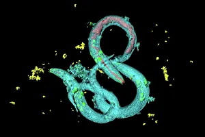
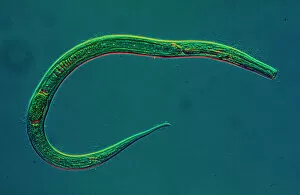
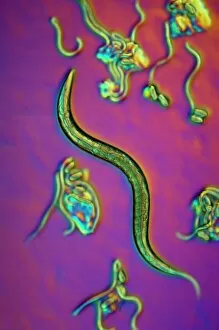
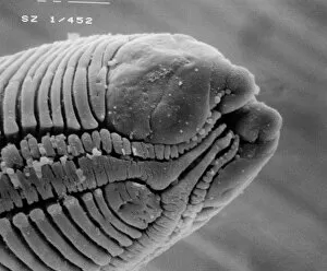
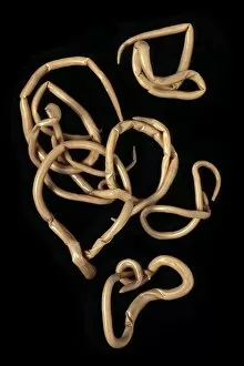
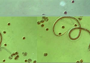
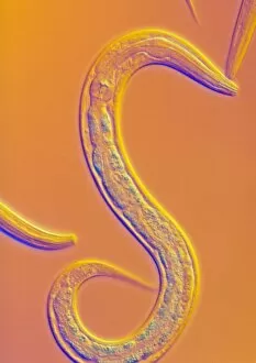
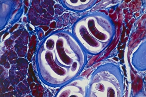
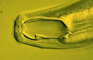
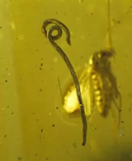
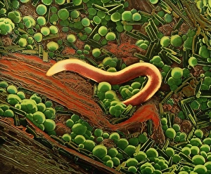
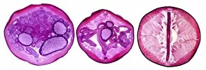
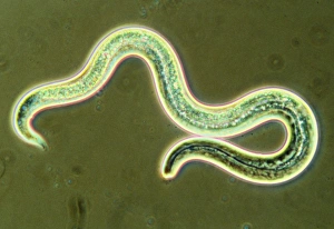
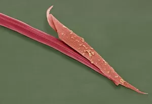
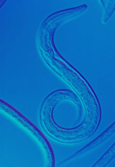
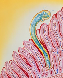
Nematoda, commonly known as nematode worms, are a diverse group of organisms that inhabit various environments worldwide. One well-studied species is C. Elegans, which has been extensively used in scientific research due to its transparent body and simple nervous system. In light micrographs of C. Elegans worms, their slender bodies can be observed with remarkable detail. These microscopic images reveal the intricate structure and organization within these tiny creatures. Another fascinating aspect captured by scanning electron microscopy (SEM) is the parasitic nature of some nematodes. Parasitic nematode worms can be seen clinging onto their hosts, showcasing their ability to adapt and survive in different ecosystems. The study of nematodes dates back centuries when Karl Rudolphi, a Swedish naturalist, first identified them as distinct organisms in the early 19th century. Since then, researchers have made significant progress in understanding these roundworms' biology and impact on human health. One such example is Ascaris lumbricoides - a human roundworm that infects millions worldwide. Light micrographs provide insights into this parasite's life cycle and its presence within the human body. Trichina larvae encysted in muscle tissue also offer captivating visuals under light microscopy. These images depict how these tiny parasites invade muscle cells and form cysts as part of their life cycle. Overall, studying nematodes provides valuable knowledge about fundamental biological processes while shedding light on the complex interactions between parasites and their hosts.