
Nematode Collection
Nematodes, commonly known as nematode worms, are fascinating creatures that have captured the attention of scientists and researchers for centuries


All Professionally Made to Order for Quick Shipping
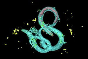
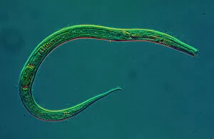
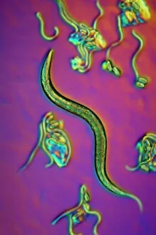
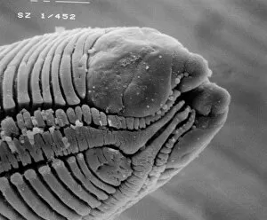
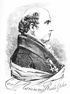
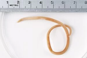
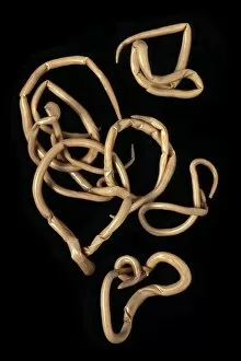
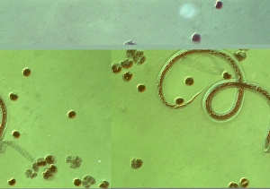
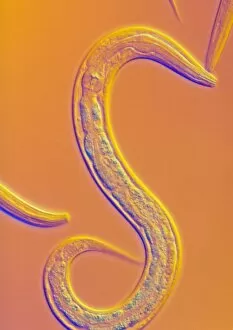
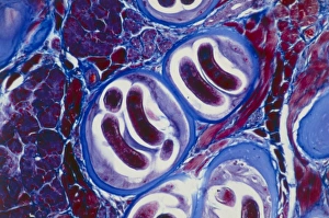
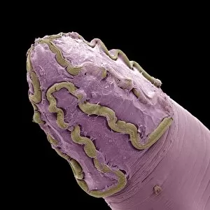
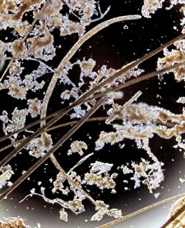
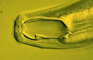
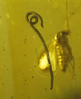
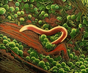
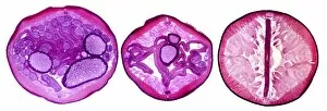
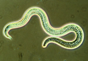
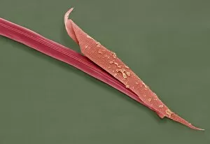
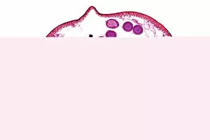
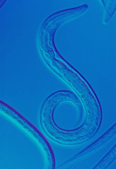
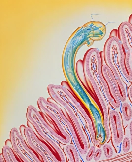
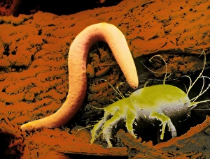
Nematodes, commonly known as nematode worms, are fascinating creatures that have captured the attention of scientists and researchers for centuries. One such species is the C. Elegans worm, which has become a model organism in biological research due to its simplicity and genetic tractability. In light micrographs of C. Elegans worms, their slender bodies can be seen wriggling gracefully through their environment. These images reveal intricate details of their anatomy and behavior, offering insights into their biology. SEM (scanning electron microscopy) images provide even more stunning views worms. The high-resolution pictures showcase the fine structure of these organisms with incredible clarity. From the rounded heads to the tapered tails, every aspect is beautifully displayed. While some nematodes like C. Elegans are harmless soil-dwelling organisms, others are parasitic and cause diseases in humans and animals alike. Parasitic nematode worms can infect various organs within their hosts' bodies, leading to severe health issues if left untreated. One well-known example is Ascaris lumbricoides – a human roundworm that affects millions worldwide. Its presence inside the intestines can lead to malnutrition and impaired growth in children while causing discomfort for adults. The study of nematodes dates back centuries when Karl Rudolphi, a Swedish naturalist, first described them in detail during his scientific endeavors. Since then, countless researchers have dedicated themselves to unraveling the mysteries surrounding these remarkable creatures. Whether it's exploring their genetics or understanding how they interact with other organisms in ecosystems, studying nematodes continues to yield valuable knowledge about life on Earth. So next time you come across an image or mention of a nematode worm like C. elegans or Ascaris lumbricoides remember that there's much more than meets the eye – from microscopic wonders under light micrographs to complex relationships between parasites and hosts – these tiny creatures hold a wealth of information waiting to be discovered.