Fraud Blocker Skip to main content

Ovum Collection (page 3)

The miracle of life begins with the ovum, a tiny but powerful entity that holds the key to creation

Background imageOvum Collection: Fertilisation, artwork C013 / 4643

Fertilisation, artwork C013 / 4643
Fertilisation. Computer artwork of numerous sperm cells (spermatozoa) swimming towards an egg (bright) in the distance

Background imageOvum Collection: Fertilisation, artwork C013 / 4644

Fertilisation, artwork C013 / 4644
Fertilisation. Computer artwork of numerous sperm cells (spermatozoa) swimming towards an egg (bright) in the distance

Background imageOvum Collection: Large Yellow Underwing (Noctua pronuba) egg batch, laid on buttercup stalk in meadow, Powys

Large Yellow Underwing (Noctua pronuba) egg batch, laid on buttercup stalk in meadow, Powys, Wales, july

Background imageOvum Collection: Liver flukes, transverse sections

Liver flukes, transverse sections
Liver flukes. Light micrographs of transverse sections through the male (top) and female (bottom) body areas of an adult liver fluke (Fasciola hepatica)

Background imageOvum Collection: Liver fluke, transverse section

Liver fluke, transverse section
Liver fluke. Light micrograph of a transverse section through the anterior half of a liver fluke (Fasciola hepatica), a hermaphrodite tremotode worm and endoparasite that infects sheep

Background imageOvum Collection: Nematode worm, transverse sections

Nematode worm, transverse sections
Nematode worm. Light micrograph of three transverse sections through the body of a female round unsegmented worm (Ascaris lumbricoides)

Background imageOvum Collection: Cubic close-packed crystal structure

Cubic close-packed crystal structure
Haemoglobin molecule. Computer artwork showing the structure of a haemoglobin molecule. Haemoglobin is a metalloprotein that transports oxygen around the body in red blood cells

Background imageOvum Collection: Butterfly eggs, SEM

Butterfly eggs, SEM
Butterfly eggs. Coloured scanning electron micrograph (SEM) of the eggs of a butterfly (order Lepidoptera). One of the eggs at lower right is hatching

Background imageOvum Collection: Broadclub cuttlefish egg

Broadclub cuttlefish egg. Egg laid by a broadclub cuttlefish (Sepia latimanus) in fire coral. Photographed near Indonesia

Background imageOvum Collection: LM of human sperm fertilising an egg

LM of human sperm fertilising an egg
Sperm fertilising egg. Light micrograph of a human egg with sperm at its surface attempting to penetrate and fertilise it

Background imageOvum Collection: Egg cell, SEM

Egg cell, SEM
Egg cell. Coloured scanning electron micrograph (SEM) of a freeze-fracture of a developing egg cell in a secondary follicle of the ovary. At centre is the rounded egg cell or secondary oocyte (red)

Background imageOvum Collection: Coloured SEM of egg cell in secondary follicle

Coloured SEM of egg cell in secondary follicle

Background imageOvum Collection: Coloured SEM of sperm fertilising an egg

Coloured SEM of sperm fertilising an egg
Coloured scanning electron micrograph of sperm (yellow) fertilising an egg cell (round and pink). At top centre three polar bodies (purple and blue) are seen

Background imageOvum Collection: Gene transfer in fertilisation

Gene transfer in fertilisation

Background imageOvum Collection: Computer artwork of human sperm fertilising an egg

Computer artwork of human sperm fertilising an egg
Sperm fertilising egg. Computer illustration of sperm cells (white) approaching a human egg (pink), before one of the sperm fertilises it

Background imageOvum Collection: Artwork of human fertilisation and implantation

Artwork of human fertilisation and implantation
Fertilisation and implantation. Artwork of the first week of human life, from egg fertilisation to embryo implantation in the uterus (womb)

Background imageOvum Collection: Ovulation, artwork

Ovulation, artwork
Ovulation. Computer artwork of an egg (yellow) being released from an ovary (bottom), a process known as ovulation. The egg is being released into a fallopian tube (top)

Background imageOvum Collection: False-colour SEM of the ovary surface at ovulation

False-colour SEM of the ovary surface at ovulation

Background imageOvum Collection: Coloured LM of a fertilised human egg 24 hours old

Coloured LM of a fertilised human egg 24 hours old

Background imageOvum Collection: False-colour SEM of secondary oocyte & polar body

False-colour SEM of secondary oocyte & polar body
Human egg. False-colour scanning electron micrograph of a human egg (primary oocyte) at the end of the first meiotic division, occurring just before ovulation

Background imageOvum Collection: SEM of ovulation; release of egg

SEM of ovulation; release of egg

Background imageOvum Collection: Egg fertilisation

Egg fertilisation
False-colour transmission electron micrograph (TEM) of a single human ovum (egg) surrounded by sperm. The egg has been coloured orange, and the numerous sperm appear rougly spherical in this section

Background imageOvum Collection: Sperm fertilising egg

Sperm fertilising egg

Background imageOvum Collection: Human fertilisation

Human fertilisation
Sperm fertilising egg. Computer illustration of sperm cells (at right) approaching a human egg (at left). One of the sperm is fertilising it

Background imageOvum Collection: Coloured SEM showing the ovulation process

Coloured SEM showing the ovulation process
Ovulation. Coloured scanning electron micrograph of the ovulation process. The egg (red at centre) has ruptured the external surface of the ovary (dark green), known as the germinal epithelium

Background imageOvum Collection: Artwork of a human egg cell

Artwork of a human egg cell in the primordial follicle phase which occurs after seven months of fetal development. The egg cell is surrounded by a thick layer of follicular cells known as zona

Background imageOvum Collection: False-colour SEM of human egg after ovulation

False-colour SEM of human egg after ovulation
Ovarys surface at ovulation. False-colour scanning electron micrograph of the surface of the ovary about one hour after ovulation

Background imageOvum Collection: Lampbrush chromosomes, TEM

Lampbrush chromosomes, TEM
Lampbrush chromosomes. Coloured transmission electron micrograph (TEM) of lampbrush chromosomes (LBCs). A chromosome consists of proteins and DNA (deoxyribonucleic acid)

Background imageOvum Collection: LM of immature human oocyte with germinal vesicle

LM of immature human oocyte with germinal vesicle
Light micrograph of an immature human oocyte (the female egg cell) containing a germinal body, the large cell nucleus. Oocytes are formed in the ovaries

Background imageOvum Collection: Follicles in a human ovary, micrograph

Follicles in a human ovary, micrograph
Developing follicles in a human ovary, light micrograph. Three developing Graafian follicles are seen. A Graafian follicle is a structure in the ovaries from which an ovum (egg cell) is released

Background imageOvum Collection: Artwork of ovum (egg) development in womans ovary

Artwork of ovum (egg) development in womans ovary
Ovum development. Illustration showing the development of a human ovum (egg) in an ovary (pink, at lower centre) during the menstrual cycle

Background imageOvum Collection: Graafian follicles, light micrograph

Graafian follicles, light micrograph
Developing Graafian follicles. Light micrograph of a sectioned human ovary, showing two developing Graafian follicles. A Graafian follicle is a structure in the ovaries from which an ovum (egg cell)

Background imageOvum Collection: Internet relationship

Internet relationship. Conceptual computer artwork of X-rays of a computer mouse representing a sperm fertilising an egg. This could represent the popularity of online dating

Background imageOvum Collection: Medical nanorobot on sperm cell

Medical nanorobot on sperm cell
Medical nanorobot. Computer artwork of a medical nanorobot holding a sperm cell. Microscopic robot technology could be developed in the future to treat disorders, such as infertility, in new ways

Background imageOvum Collection: Sperm fertilising an egg, artwork

Sperm fertilising an egg, artwork
Sperm fertilising an egg. Cutaway artwork of a human sperm cell (spermatozoon) penetrating an egg cells (ovum) thick outer layer (zona pellucida). The egg cells membrane is at top right

Background imageOvum Collection: Fertilisation, computer artwork

Fertilisation, computer artwork
Fertilisation. Computer artwork of a sperm cell (purple) penetrating a human egg (ovum). Each sperm (spermatozoan) has a rounded head (acrosome) and a long tail with which it swims

Background imageOvum Collection: Fertilisation

Fertilisation, computer artwork. Sperm fertilising an egg

Background imageOvum Collection: Fertilization

Fertilization. Artwork of sperm cells (white and black) surrounding a human egg cell (round). The egg cell is protected by an outer layer, the zona pellucida (black)

Background imageOvum Collection: Egg, SEM

Egg, SEM
Egg. Coloured scanning electron micrograph (SEM) of a type of egg. This egg was found on the back of a common shore crab (Carcinus maenas)

Background imageOvum Collection: Ovarian follicles, light micrograph

Ovarian follicles, light micrograph
Ovarian follicles. Coloured light micrograph of a section through an ovary (orange), showing three primary follicles (pink)


All Professionally Made to Order for Quick Shipping

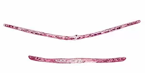
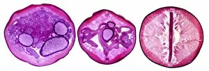
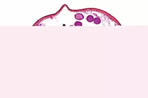
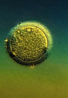
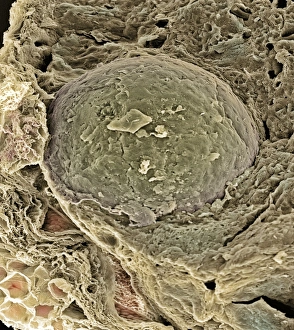
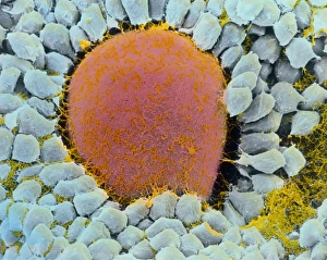
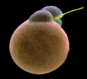
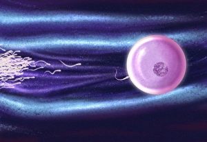
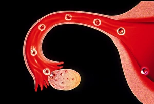
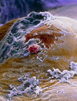
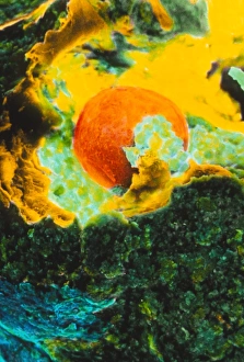
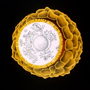
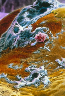
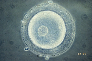
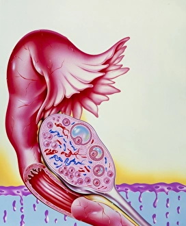
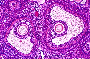
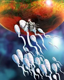
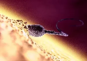
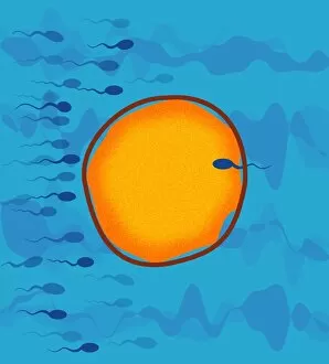
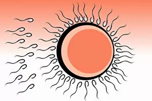
The miracle of life begins with the ovum, a tiny but powerful entity that holds the key to creation. In this captivating coloured scanning electron microscope (SEM) image, we witness the intricate dance between an egg and sperm during fertilisation. Picture No. 11675528 reveals the mesmerizing beauty of this process as we zoom in on the moment when life takes its first breath. The delicate structure of the ovum is highlighted, showcasing its remarkable ability to attract and unite with a single sperm among millions. As we delve deeper into Picture No. 11675527, our eyes are drawn to the vibrant hues that illuminate this microscopic world, and is here that we witness nature's masterpiece unfolding before us – an extraordinary fusion of two distinct entities merging into one. In Picture No. 11675526, our focus shifts towards understanding the origin of these incredible cells within a woman's body. The ovarian follicle takes center stage in this SEM image, reminding us of the complex journey each ovum undertakes before it reaches maturity. Stepping back in time through Picture No. 11675525, we encounter Karl Ernst von Baer – a visionary biologist whose groundbreaking work shaped our understanding of embryology and reproduction. His illustrations from "La Creation Naturelle et les Etres" depict human fertilisation with astonishing accuracy and detail. Karl Ernst von Baer himself comes to life in Picture No. 11675522 – his portrait capturing both his brilliance as a scientist and his unwavering dedication to unraveling nature's mysteries. Finally, an illustration from another era transports us back to ancient times when humans marveled at conception's wonders depicted by Karl Ernst von Baer (1792-1876). This timeless artwork serves as a testament to humanity's eternal fascination with new beginnings and our ceaseless quest for knowledge about life itself. These captivating images offer glimpses into different facets surrounding ovum and fertilisation.