Fraud Blocker Skip to main content

Parasitology Collection (page 2)

"Exploring the Intricate World of Parasitology: Unveiling Nature's Hidden Players" Delving into the Microcosm

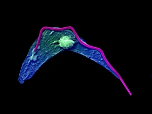
Background imageParasitology Collection: Cytoskeleton in unicellular parasite, SEM C018 / 0518

Cytoskeleton in unicellular parasite, SEM C018 / 0518
Cytoskeleton in unicellular parasite, coloured scanning electron micrograph (SEM). All cells have a support and transport network called the cytoskeleton

Background imageParasitology Collection: Malaria infection cycle, artwork

Malaria infection cycle, artwork. Life cycle of the Plasmodium parasite that causes malaria, which can cause coma and death. At upper left, a female Anopheles mosquito bites and infects a person

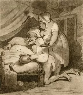
Background imageParasitology Collection: Catching bedbugs, 18th century

Catching bedbugs, 18th century
Catching bedbugs. 18th-century artwork, titled Summer Amusement - Bugg Hunting, showing an elderly couple catching bedbugs

Background imageParasitology Collection: Tick on a snake C014 / 0957

Tick on a snake C014 / 0957
Tick on a snake. Parasitic tick (superfamily Ixodoidea, white) embedded in the body of Waglers sipo, or smooth machete savane, (Chironius scurrulus) snake

Background imageParasitology Collection: Parasitic fungus on a weevil C014 / 0951

Parasitic fungus on a weevil C014 / 0951
Parasitic fungus on a weevil. Fruiting body of a Cordyceps sp. fungus (left) growing out of the body of a weevil (superfamily Curculionoidea, right) clinging to a plant stem

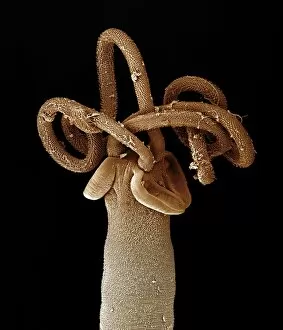
Background imageParasitology Collection: Tapeworm head, SEM C014 / 4877

Tapeworm head, SEM C014 / 4877
Tapeworm head. Coloured scanning electron micrograph of the head (scolex) of a tapeworm (class Cestoda) found in a shark, showing the hooks used to attach to the host animals digestive tract

Background imageParasitology Collection: Tapeworm head, SEM C014 / 4880

Tapeworm head, SEM C014 / 4880
Tapeworm head. Coloured scanning electron micrograph of the head (scolex) of a tapeworm (Platybothrium auriculatum) found in a blue shark (Prionace glauca)

Background imageParasitology Collection: Tapeworm head, SEM C014 / 4878

Tapeworm head, SEM C014 / 4878
Tapeworm head. Coloured scanning electron micrograph of the head (scolex) of a tapeworm (class Cestoda) found in a shark, showing the hooks used to attach to the host animals digestive tract

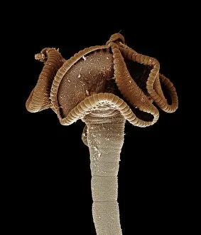
Background imageParasitology Collection: Tapeworm head, SEM C014 / 4879

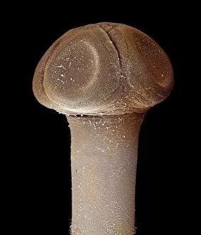
Tapeworm head, SEM C014 / 4879
Tapeworm head. Coloured scanning electron micrograph of the head (scolex) of a tapeworm (class Cestoda) found in a shark, showing the suckers used to attach to the host animals digestive tract

Background imageParasitology Collection: Honey bee mite, SEM C016 / 8024

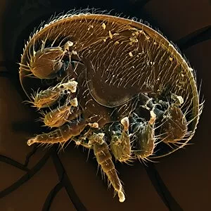
Honey bee mite, SEM C016 / 8024
Honey bee mite. Coloured scanning electron micrograph (SEM) of the underside of a Varroa sp. mite from a honey bee. Varroa are parasitic mites that attack honey bees (Apis sp.)

Background imageParasitology Collection: Anopheles stephensi mosquito larva

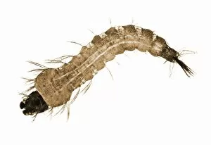
Anopheles stephensi mosquito larva. The head is at left, the tail at right. The adult Anopheles stephensi female is a known vector for the tropical disease malaria

Background imageParasitology Collection: Malarial blood cell, SEM C017 / 8307

Malarial blood cell, SEM C017 / 8307
Malarial blood cell. Coloured scanning electron micrograph (SEM) of an erythrocyte (red blood cell) infected with Plasmodium parasites, the cause of malaria

Background imageParasitology Collection: Veterinarians Theiler and Mohler in 1923 C017 / 7824

Veterinarians Theiler and Mohler in 1923 C017 / 7824
Veterinarians Theiler and Mohler in 1923. Swiss-South African veterinarian Arnold Theiler (left, 1867-1936) and US veterinarian John Robbins Mohler (right, 1875-1952)

Background imageParasitology Collection: Raphael Blanchard, French parasitologist C016 / 5114

Raphael Blanchard, French parasitologist C016 / 5114
Raphael Blanchard (1857-1919), French physician and parasitologist. Blanchards positions included Professor at the Paris Faculty of Medicine

Background imageParasitology Collection: Zombie ant fungus life-cycle, artwork

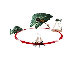
Zombie ant fungus life-cycle, artwork
Zombie ant fungus life-cycle. Computer artwork showing the life cycle of the parasitoidal fungus Ophiocordyceps unilateralis

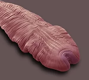
Background imageParasitology Collection: Tapeworm head, SEM C016 / 3093

Tapeworm head, SEM C016 / 3093
Tapeworm head. Coloured scanning electron micrograph (SEM) of the head of a tapeworm (class Cestoda) in the cysticercoid stage

Background imageParasitology Collection: Tapeworm head, SEM C016 / 3092

Tapeworm head, SEM C016 / 3092
Tapeworm head. Coloured scanning electron micrograph (SEM) of the head of a tapeworm (class Cestoda) in the cysticercoid stage

Background imageParasitology Collection: Gapeworm, SEM C016 / 3085

Gapeworm, SEM C016 / 3085
Gapeworm. Coloured scanning electron micrograph of a female gapeworm (Syngamus trachea) from an owl. Gapeworms are parasitic nematode worms that infect the trachea (windpipe) of certain birds

Background imageParasitology Collection: Gapeworm, SEM C016 / 3084

Gapeworm, SEM C016 / 3084
Gapeworm. Coloured scanning electron micrograph of a female gapeworm (Syngamus trachea) from an owl. Gapeworms are parasitic nematode worms that infect the trachea (windpipe) of certain birds

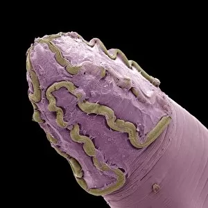
Background imageParasitology Collection: Gapeworm head, SEM C016 / 3082

Gapeworm head, SEM C016 / 3082
Gapeworm head. Coloured scanning electron micrograph of the head of a female gapeworm (Syngamus trachea) from an owl. Gapeworms are parasitic nematode worms that infect the trachea (windpipe)

Background imageParasitology Collection: Gapeworm head, SEM C016 / 3083

Gapeworm head, SEM C016 / 3083
Gapeworm head. Coloured scanning electron micrograph of the head of a female gapeworm (Syngamus trachea) from an owl. Gapeworms are parasitic nematode worms that infect the trachea (windpipe)

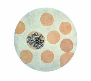
Background imageParasitology Collection: Malaria parasite in red blood cell C016 / 2444

Malaria parasite in red blood cell C016 / 2444
Malaria parasite in red blood cell. Light micrograph of a red blood cell (erythrocyte) infected with a Plasmodim vivax protozoan. This parasite causes recurring malaria in humans

Background imageParasitology Collection: Malaria parasite in red blood cell C016 / 2443

Malaria parasite in red blood cell C016 / 2443
Malaria parasite in red blood cell. Light micrograph of a red blood cell (erythrocyte) infected with a Plasmodim vivax protozoan. This parasite causes recurring malaria in humans

Background imageParasitology Collection: Malaria parasite in red blood cell C016 / 2442

Malaria parasite in red blood cell C016 / 2442
Malaria parasite in red blood cell. Light micrograph of a red blood cell (erythrocyte) infected with a Plasmodim vivax protozoan. This parasite causes recurring malaria in humans

Background imageParasitology Collection: Human head louse Pediculus, LM C013 / 5282

Human head louse Pediculus, LM C013 / 5282
Light micrograph using differential interference contrast (DIC) of a human head louse (Pediculus humanus capitus). This exoparasite has a large head with two eyes (black)

Background imageParasitology Collection: Nematode worms, SEM

Nematode worms, SEM
Nematode worm. Scanning electron micrograph of a juvenile Heterorhabditis bacteriophora nematode worm (centre) shedding its cuticle (outer coat, centre right) as it develops into an adult

Background imageParasitology Collection: Head lice, artwork

Head lice, artwork
Head lice (Pediculus humanus capitis), computer artwork. The louse at left has its lower (ventral) side facing upwards and the louse at right has its upper (dorsal) side facing upwards

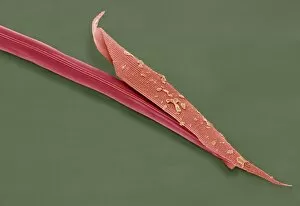
Background imageParasitology Collection: Squirrel flea antenna, SEM

Squirrel flea antenna, SEM
Squirrel flea antenna. Coloured scanning electron micrograph (SEM) of the antenna of a Squirrel flea (Orchopeas howardi howardi)

Background imageParasitology Collection: Squirrel flea, SEM

Squirrel flea, SEM
Squirrel flea (Orchopeas howardi howardi), coloured scanning electron micrograph (SEM). The two structures protruding from the centre of the fleas head are sensory palps

Background imageParasitology Collection: Malaria, conceptual artwork

Malaria, conceptual artwork. Drop of red blood, showing the outline of the continent of Africa. Malaria is widespread in tropical and subtropical regions, including parts of the Americas, Asia

Background imageParasitology Collection: Bilharzia infection, ureter tissue

Bilharzia infection, ureter tissue
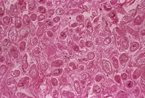
Bilharzia infection. Light micrograph of human ureter tissue that contains numerous eggs from Schistosoma flukes. The ureters are tubes that carry urine from the kidneys to the bladder

Background imageParasitology Collection: Addison Emery Verrill, US zoologist

Addison Emery Verrill, US zoologist
Addison Emery Verrill (1839-1926), American zoologist. Verrill graduated from Harvard University, USA, but moved to Yale University when he was appointed as the first professor of zoology there

Background imageParasitology Collection: Tick feeding, artwork

Tick feeding, artwork

Background imageParasitology Collection: Eyelash mites, artwork

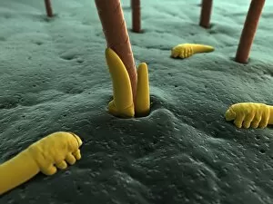
Eyelash mites, artwork

Background imageParasitology Collection: Eyelash mite, artwork

Eyelash mite, artwork

Background imageParasitology Collection: Cat flea, artwork

Cat flea, artwork


All Professionally Made to Order for Quick Shipping

"Exploring the Intricate World of Parasitology: Unveiling Nature's Hidden Players" Delving into the Microcosm: A mesmerizing artwork captures the elusive Trypanosome protozoan, unraveling its mysterious existence in parasitology. Illuminating Secrets: Witness the intricate details of an Anopheles mosquito male through a captivating light micrograph, shedding light on its role as a carrier of deadly parasites. Unraveling Nature's Enigma: Dive deep into the microscopic realm with stunning SEM images showcasing nematode worms, revealing their complex adaptations and survival strategies. Pioneers of Knowledge: Meet Saul Adler (1895-1966), a Russian-born British medical scientist whose groundbreaking research revolutionized our understanding of parasitology. Trailblazers in Discovery: Discover Karl Rudolphi, a Swedish naturalist who made significant contributions to early studies on parasites, paving the way for future advancements in this field. Timeless Artistry: Journey back in time with historical artwork depicting tapeworms, providing glimpses into how these parasites have captivated human imagination throughout history. The Battle Within: Experience a conceptual image illustrating plasmodium causing malaria - an ongoing war between humans and these cunning parasites that claim countless lives worldwide. Bloodborne Threats Unveiled: Witness African trypanosomiasis lurking within red blood cells through striking visuals that highlight both the beauty and danger posed by these microscopic invaders. Dance of Destruction: Observe another conceptual image portraying Trypanosoma - tiny yet formidable organisms capable of wreaking havoc within their unsuspecting hosts' bodies. Silent Invaders Revealed: Peer inside red blood cells with awe-inspiring conceptual imagery exposing malaria parasites at work - stealthy intruders responsible for one of humanity's deadliest diseases.