Fraud Blocker Skip to main content

Parasitology Collection (page 5)

"Exploring the Intricate World of Parasitology: Unveiling Nature's Hidden Players" Delving into the Microcosm

Background imageParasitology Collection: Head louse, light micrograph

Head louse, light micrograph

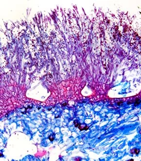
Background imageParasitology Collection: Penicillium mould, light micrograph

Penicillium mould, light micrograph
Penicillium mould. Light micrograph of a section through an orange rind infected with Penicillium sp. blue mould fungus, showing fungal hyphae and conidiophores (pink and purple) with spores (red)

Background imageParasitology Collection: Beef liver fluke, light micrograph

Beef liver fluke, light micrograph

Background imageParasitology Collection: Downy mildew infection, light micrograph

Downy mildew infection, light micrograph
Downy mildew infection. Light micrograph of a section through a grapevine (Vitis sp.) leaf infected with downy mildew (Plasmopara viticola)

Background imageParasitology Collection: Head of a human flea, light micrograph

Head of a human flea, light micrograph

Background imageParasitology Collection: Hollyhock rust fungus in a leaf

Hollyhock rust fungus in a leaf. Light micrograph of a section through a hollyhock (Alcea sp.) leaf infected with the parasitic Puccinia malvacearum rust, showing the teliospores (spores)

Background imageParasitology Collection: Clubroot infection, light micrograph

Clubroot infection, light micrograph
Clubroot infection. Light micrograph a section through a cabbage root infected with clubroot (Plasmodiophora brassicae). Plasmodiophora is a parasitic protist that infects the roots of cabbages

Background imageParasitology Collection: Chicken mite, light micrograph

Chicken mite, light micrograph

Background imageParasitology Collection: Testing ticks in public health lab

Testing ticks in public health lab
Public health. A researcher gathering samples of ticks in a public health laboratory. Ticks (superfamily Ixodoidea) are parasites that eat the blood of their hosts

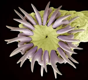
Background imageParasitology Collection: Tapeworm head, SEM

Tapeworm head, SEM
Tapeworm head. Coloured scanning electron micrograph of the head (scolex) of a tapeworm (Taenia pisiformis), showing the hooks used to attach to the host animals digestive tract

Background imageParasitology Collection: Parasite and cells

Parasite and cells
Concept image. An aggresive unknown Parasite has entered a cell and consumes it from within

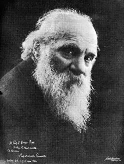
Background imageParasitology Collection: Edoardo Perroncito, Italian physician

Edoardo Perroncito, Italian physician
Edoardo Perroncito (1847Ôé¼" 1936), Italian physician, veterinarian and parasitologist. Perroncito is credited, along with Luigi Pagliani (1847Ôé¼" 1931)

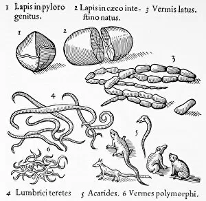
Background imageParasitology Collection: Human parasites, historical artwork

Human parasites, historical artwork
Human parasites. 1575 artwork of human parasites drawn by the Dutch physician, astronomer and astrologer Cornelius Gemma (1535Ôé¼" 1579)

Background imageParasitology Collection: Giovanni Grassi, Italian zoologist

Giovanni Grassi, Italian zoologist
Giovanni Grassi. Portrait of Giovanni Batista Grassi (1854-1925), Italian zoologist and malaria pioneer. In 1889-90 Grassi suggested that different species of the Plasmodium protozoa caused different

Background imageParasitology Collection: Intestinal parasites, artwork

Intestinal parasites, artwork
Intestinal parasites, computer artwork. Parasitic worms embedded in the lining of the intestine. A number of worms, including threadworm, hookworm and tapeworm, are parasitic on the human gut

Background imageParasitology Collection: Mosquito testis infected with bacteria

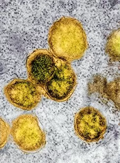
Mosquito testis infected with bacteria. Coloured transmission electron micrograph (TEM) of a testis from a Culex pipiens mosquito infected with Walbachia sp. bacteria (yellow)

Background imageParasitology Collection: Mosquito ovary infected with bacteria TEM

Mosquito ovary infected with bacteria TEM
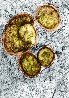
Mosquito ovary infected with bacteria. Coloured transmission electron micrograph (TEM) of an ovary from a Culex pipiens mosquito infected with Walbachia sp. bacteria (yellow)

Background imageParasitology Collection: Artichoke Galls on Pedunculate Oak

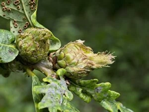
Artichoke Galls on Pedunculate Oak (Quercus rubus). Flower buds infected by the gall wasp Andricus fecundator. In spring the female gall wasp lays single eggs in buds of the Pedunculate oak

Background imageParasitology Collection: Red blood cells with malaria, artwork

Red blood cells with malaria, artwork
Red blood cells with malaria, computer artwork. Malaria is caused by the Plasmodium parasite, which is transmitted into the bloodstream by mosquito bites

Background imageParasitology Collection: Dodder parasitic plant

Dodder parasitic plant. Polarised light micrograph of a section through a host plant parasitised by a dodder (Cuscuta sp.)

Background imageParasitology Collection: Plant parasite, light micrograph

Plant parasite, light micrograph
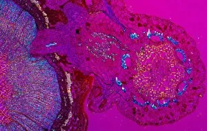
Plant parasite. Polarised light micrograph of a transverse section through a plant stem (left) entwined by the common dodder (Cuscuta sp.) parasitic plant (round, right)

Background imageParasitology Collection: Mange mites, SEM

Mange mites, SEM
Mange mites. Coloured scanning electron micrograph (SEM) of two mites of different sizes belonging to the family Sarcoptidae

Background imageParasitology Collection: Mange mite, SEM

Mange mite, SEM
Mange mite. Coloured scanning electron micrograph (SEM) of a mite belonging to the family Sarcoptidae. It is crawling through dog hair


All Professionally Made to Order for Quick Shipping

"Exploring the Intricate World of Parasitology: Unveiling Nature's Hidden Players" Delving into the Microcosm: A mesmerizing artwork captures the elusive Trypanosome protozoan, unraveling its mysterious existence in parasitology. Illuminating Secrets: Witness the intricate details of an Anopheles mosquito male through a captivating light micrograph, shedding light on its role as a carrier of deadly parasites. Unraveling Nature's Enigma: Dive deep into the microscopic realm with stunning SEM images showcasing nematode worms, revealing their complex adaptations and survival strategies. Pioneers of Knowledge: Meet Saul Adler (1895-1966), a Russian-born British medical scientist whose groundbreaking research revolutionized our understanding of parasitology. Trailblazers in Discovery: Discover Karl Rudolphi, a Swedish naturalist who made significant contributions to early studies on parasites, paving the way for future advancements in this field. Timeless Artistry: Journey back in time with historical artwork depicting tapeworms, providing glimpses into how these parasites have captivated human imagination throughout history. The Battle Within: Experience a conceptual image illustrating plasmodium causing malaria - an ongoing war between humans and these cunning parasites that claim countless lives worldwide. Bloodborne Threats Unveiled: Witness African trypanosomiasis lurking within red blood cells through striking visuals that highlight both the beauty and danger posed by these microscopic invaders. Dance of Destruction: Observe another conceptual image portraying Trypanosoma - tiny yet formidable organisms capable of wreaking havoc within their unsuspecting hosts' bodies. Silent Invaders Revealed: Peer inside red blood cells with awe-inspiring conceptual imagery exposing malaria parasites at work - stealthy intruders responsible for one of humanity's deadliest diseases.