
Parasitology Collection
"Exploring the Intricate World of Parasitology: Unveiling Nature's Hidden Players" Delving into the Microcosm


All Professionally Made to Order for Quick Shipping
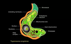
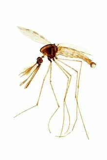
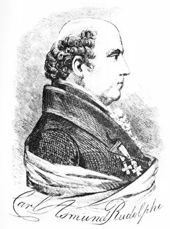
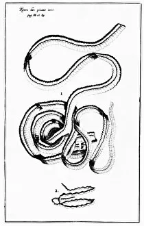
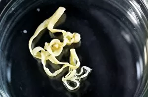
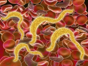
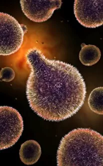
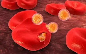
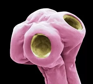
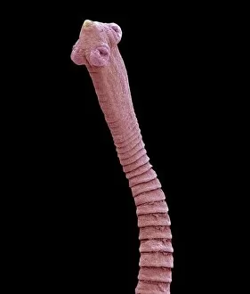
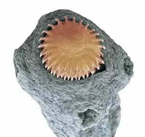
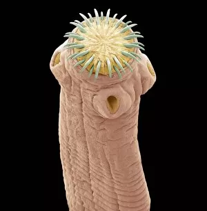
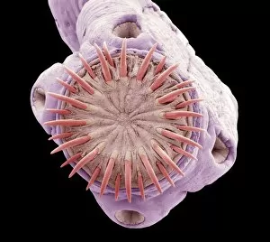
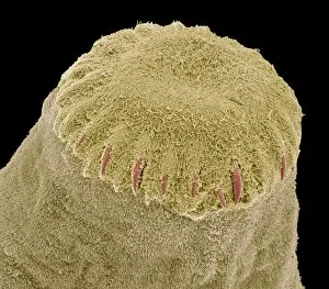
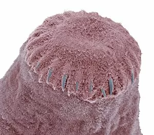
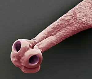
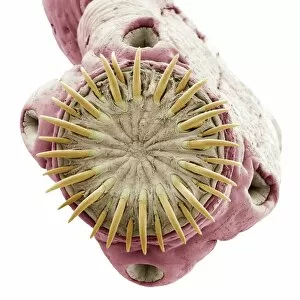
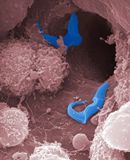
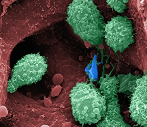
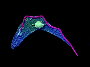
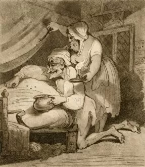
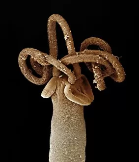
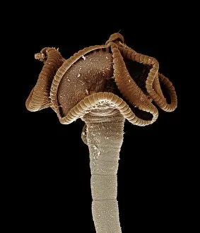
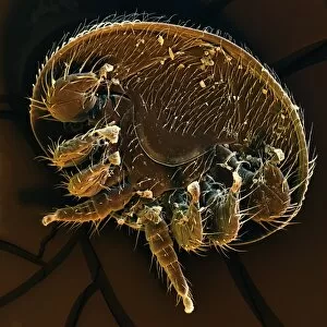
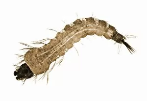
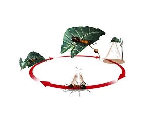
"Exploring the Intricate World of Parasitology: Unveiling Nature's Hidden Players" Delving into the Microcosm: A mesmerizing artwork captures the elusive Trypanosome protozoan, unraveling its mysterious existence in parasitology. Illuminating Secrets: Witness the intricate details of an Anopheles mosquito male through a captivating light micrograph, shedding light on its role as a carrier of deadly parasites. Unraveling Nature's Enigma: Dive deep into the microscopic realm with stunning SEM images showcasing nematode worms, revealing their complex adaptations and survival strategies. Pioneers of Knowledge: Meet Saul Adler (1895-1966), a Russian-born British medical scientist whose groundbreaking research revolutionized our understanding of parasitology. Trailblazers in Discovery: Discover Karl Rudolphi, a Swedish naturalist who made significant contributions to early studies on parasites, paving the way for future advancements in this field. Timeless Artistry: Journey back in time with historical artwork depicting tapeworms, providing glimpses into how these parasites have captivated human imagination throughout history. The Battle Within: Experience a conceptual image illustrating plasmodium causing malaria - an ongoing war between humans and these cunning parasites that claim countless lives worldwide. Bloodborne Threats Unveiled: Witness African trypanosomiasis lurking within red blood cells through striking visuals that highlight both the beauty and danger posed by these microscopic invaders. Dance of Destruction: Observe another conceptual image portraying Trypanosoma - tiny yet formidable organisms capable of wreaking havoc within their unsuspecting hosts' bodies. Silent Invaders Revealed: Peer inside red blood cells with awe-inspiring conceptual imagery exposing malaria parasites at work - stealthy intruders responsible for one of humanity's deadliest diseases.